Review
. 2021 Sep 13:12:703525.
doi: 10.3389/fmicb.2021.703525. eCollection 2021.

Energy Conservation in Fermentations of Anaerobic Bacteria

Affiliations
Review

Energy Conservation in Fermentations of Anaerobic Bacteria

Wolfgang Buckel. Front Microbiol. .

Abstract

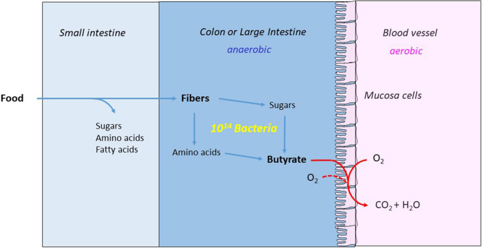
Anaerobic bacteria ferment carbohydrates and amino acids to obtain energy for growth. Due to the absence of oxygen and other inorganic electron acceptors, the substrate of a fermentation has to serve as electron donor as well as acceptor, which results in low free energies as compared to that of aerobic oxidations. Until about 10 years ago, anaerobes were thought to exclusively use substrate level phosphorylation (SLP), by which only part of the available energy could be conserved. Therefore, anaerobes were regarded as unproductive and inefficient energy conservers. The discovery of electrochemical Na+ gradients generated by biotin-dependent decarboxylations or by reduction of NAD+ with ferredoxin changed this view. Reduced ferredoxin is provided by oxidative decarboxylation of 2-oxoacids and the recently discovered flavin based electron bifurcation (FBEB). In this review, the two different fermentation pathways of glutamate to ammonia, CO2, acetate, butyrate and H2 via 3-methylaspartate or via 2-hydroxyglutarate by members of the Firmicutes are discussed as prototypical examples in which all processes characteristic for fermentations occur. Though the fermentations proceed on two entirely different pathways, the maximum theoretical amount of ATP is conserved in each pathway. The occurrence of the 3-methylaspartate pathway in clostridia from soil and the 2-hydroxyglutarate pathway in the human microbiome of the large intestine is traced back to the oxygen-sensitivity of the radical enzymes. The coenzyme B12-dependent glutamate mutase in the 3-methylaspartate pathway tolerates oxygen, whereas 2-hydroxyglutaryl-CoA dehydratase is extremely oxygen-sensitive and can only survive in the gut, where the combustion of butyrate produced by the microbiome consumes the oxygen and provides a strict anaerobic environment. Examples of coenzyme B12-dependent eliminases are given, which in the gut are replaced by simpler extremely oxygen sensitive glycyl radical enzymes.

Keywords: Rnf; coenzyme B12; decarboxylation; electron bifurcation; ferredoxin; glycyl radical enzymes; oxygen sensitivity; ΔμNa+.


Conflict of interest statement

The author declares that the research was conducted in the absence of any commercial or financial relationships that could be construed as a potential conflict of interest.

Figures

FIGURE 1
FIGURE 1
An example of energy conservation in butyrate producing anaerobic bacteria via the generation of an ion motif force. The bifurcating EtfAB-Bcd complex reduces 2 Fd with NADH driven by the exergonic reduction of crotonyl-CoA to butyryl-CoA with a second NADH. The Rnf complex in the membrane produces 2 ΔμNa+ from the exergonic reduction of NAD+ by 2 Fd. The ATPase generates 1 ATP from 4 ΔμNa+.
FIGURE 2
FIGURE 2
Partial crystal structures of the recombinant Etf-Bcd complex from C. difficile produced in E. coli; EtfAB and 2 subunits of the tetrameric Bcd are displayed. The dehydratase state shows the structure as solved. In the bifurcation state domain II of Etf rotated CW by 80° as found in Etf from A. fermentans (see red arrow); Etf, domains I + II (subunit A) in green, domain III (subunit B) in light brown, Bcd1 in yellow and Bcd2 in pinkish-brown. In the dehydratase state, α-FAD and δ-FAD are located close together (8 Å distance), ready for ET. In the bifurcation-like state, α-FAD and β-FAD are 18 Å apart; further rotation by 10° CW would bring them closer together by 4 Å, ready for electron bifurcation (taken from Buckel and Thauer, 2018a).
FIGURE 3
FIGURE 3
Scheme of electron bifurcation in the EtfAB-Bcd complex. The different FADs are placed according to their approximate reduction potential. NADH reduces β-FAD to β-FADH, which bifurcates. One electron goes to α-FAD and the formed semiquinone, β-FAD•−, reduces ferredoxin (Fd) to Fd. The formed hydroquinone (α-FADH) swings over to Bcd and transfers one electron to δ-FAD. Note the decrase of the reduction potential of α-FAD•− due to the change in location from Etf to Bcd. Repetition of this process yields δ-FADH, which reduces crotonyl-CoA to butyryl-CoA. The FAD semiquinones are red, the quinones yellow and the hydroquinones colorless (Sucharitakul et al., 2021a,b).
FIGURE 4
FIGURE 4
Fermentation of glutamate via 2-hydroxyglutarate or via 3-methylaspartate. The numbers in the circles represent the corresponding enzymes; red numbers denote radical enzymes: 1, (S)-glutamate dehydrogenase; 2, (R)-2-hydroxyglutarate dehydrogenase; 3, glutaconate CoA-transferase; 4, (R)-2-hydroxyglutaryl-CoA dehydratase; 5, glutaconyl-CoA decarboxylase, Na+-pumping; 6, glutamate mutase, coenzyme B12-dependent; 7, methylaspartase; 8, mesaconase; 9, (S)-citramalate lyase; 10, pyruvate:ferredoxin oxidoreductase (PFOR). Ac-CoA, acetyl or glutaconyl-CoA; Fd, ferredoxin; Fd, reduced ferredoxin. The formation of butyrate and acetate are shown in Figure 5. Hydrogen, H2 is formed from 2 H+ and 2 Fd, catalyzed by a [FeFe]-hydrogenase.
FIGURE 5
FIGURE 5
Butyrate synthesis in anaerobic bacteria. 1, Thiolase; 2, (S)-3-hydroxybutyryl-CoA dehydrogenases, NADH and NADPH specific; 3, (S)-3-hydroxybutyryl-CoA dehydratase; 4, electron bifurcating EtfAB-butyryl-CoA dehydrogease complex; 5, butyrate CoA-transferase.
FIGURE 6
FIGURE 6
Butyrate nourishes the mucosa cells and makes the colon anaerobic. The major part of the food is digested and absorbed in the small intestine. The remaining fibers are hydrolyzed to monosaccharides and amino acids by the anaerobic bacteria and fermented the large intestine or colon to acetate, propionate and butyrate. The mucosa cells combust the produced butyrate with oxygen from blood and colon, whereby the colon becomes strictly anaerobic.

References

    1. Anbar A. D., Duan Y., Lyons T. W., Arnold G. L., Kendall B., Creaser R. A., et al. (2007). A whiff of oxygen before the great oxidation event? Science 317 1903–1906. 10.1126/science.1140325 - DOI - PubMed
    1. Barker H. A. (1981). Amino acid degradation by anaerobic bacteria. Annu. Rev. Biochem. 50 23–40. 10.1146/annurev.bi.50.070181.000323 - DOI - PubMed
    1. Barker H. A., Kahn J. M., Hedrick L. (1982). Pathway of lysine degradation in Fusobacterium nucleatum. J. Bacteriol. 152 201–207. 10.1128/jb.152.1.201-207.1982 - DOI - PMC - PubMed
    1. Barker H. A., Smyth R. D., Wilson R. M., Weissbach H. (1959). The purification and properties of beta-methylaspartase. J. Biol. Chem. 234 320–328. 10.1016/s0021-9258(18)70297-4 - DOI - PubMed
    1. Barker H. A., Suzuki F., Iodice A., Rooze V. (1964). Glutamate mutase reaction. Ann. N. Y. Acad. Sci. 112 644–654. 10.1111/j.1749-6632.1964.tb45041.x - DOI - PubMed

LinkOut - more resources