. 2021 Apr 2;6(3):e10219.
doi: 10.1002/btm2.10219. eCollection 2021 Sep.

An engineered probiotic secreting Sj16 ameliorates colitis via Ruminococcaceae/butyrate/retinoic acid axis

Affiliations

An engineered probiotic secreting Sj16 ameliorates colitis via Ruminococcaceae/butyrate/retinoic acid axis

Lifu Wang et al. Bioeng Transl Med. .

Abstract

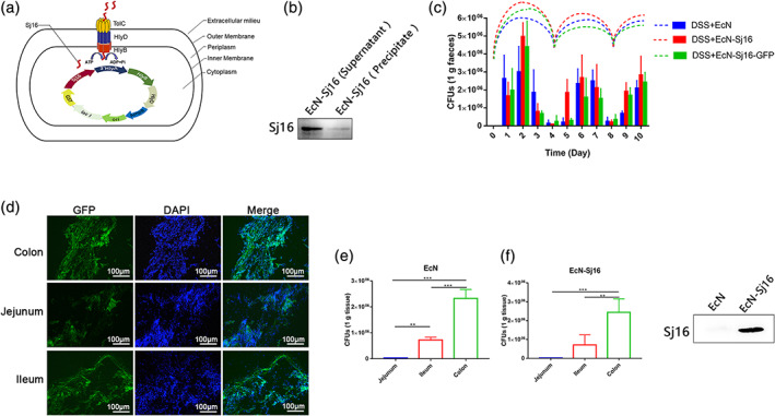
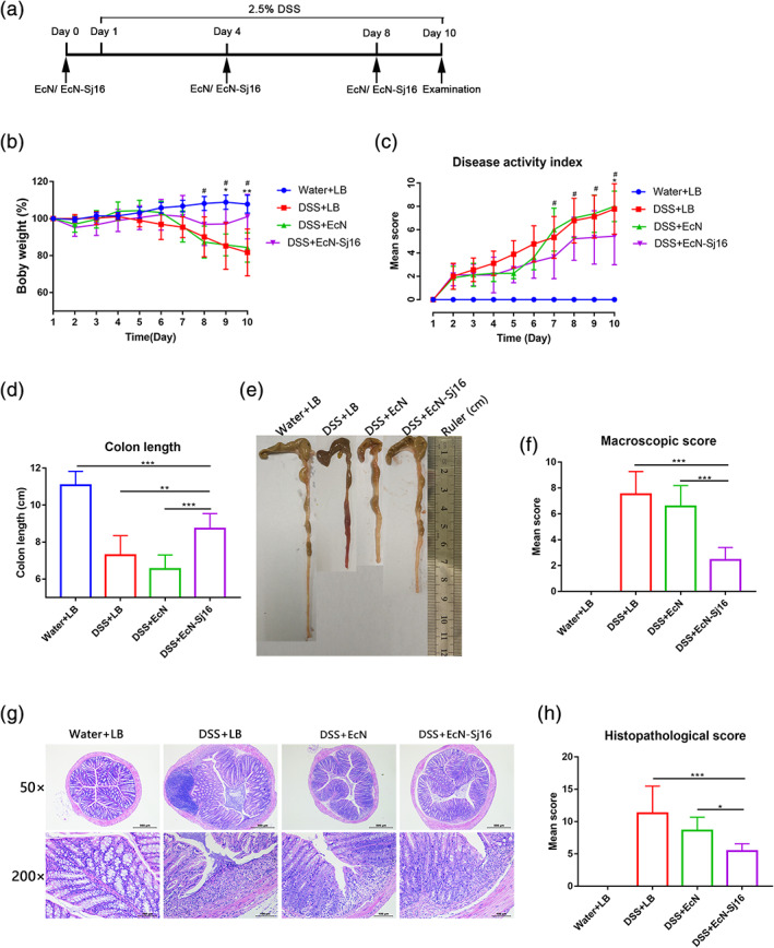
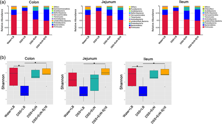
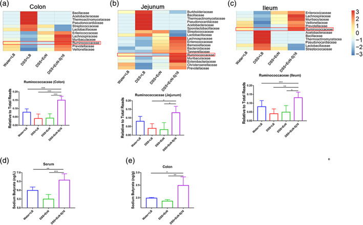
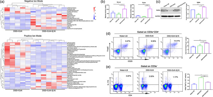
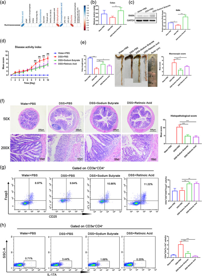
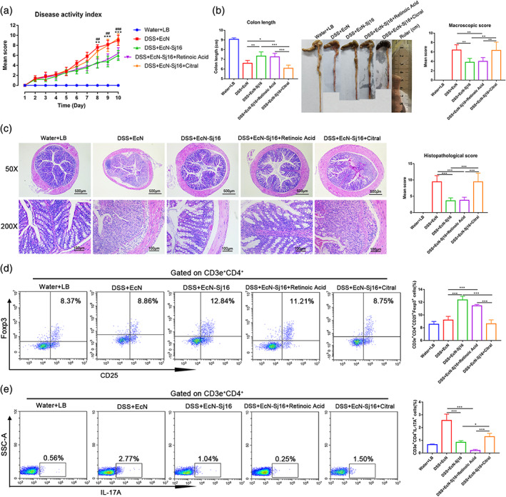
Most inflammatory bowel disease (IBD) patients are unable to maintain a lifelong remission. Developing a novel therapeutic strategy is urgently needed. In this study, we adopt a new strategy to attenuate colitis using the Escherichia coli Nissle 1917 probiotic strain to express a schistosome immunoregulatory protein (Sj16) in the gastrointestinal tract. The genetically engineered Nissle 1917 (EcN-Sj16) highly expressed Sj16 in the gastrointestinal tracts of dextran sulfate sodium-induced colitis mice and significantly attenuated the clinical activity of colitis mice. Mechanistically, EcN-Sj16 increased the intestinal microbiota diversity and selectively promoted the growth of Ruminococcaceae and therefore enhanced the butyrate production. Butyrate induced the expression of retinoic acid, which further attenuated the clinical activity of colitis mice by increasing Treg cells and decreasing Th17. Strikingly, retinoic acid inhibitor inhibited the therapeutic effects of EcN-Sj16 in colitis mice. These findings suggest that EcN-Sj16 represents a novel engineered probiotic that may be used to treat IBD.

Keywords: Ruminococcaceae/butyrate/retinoic acid axis; Sj16; Treg/Th17 balance; colitis; engineered probiotic.


Conflict of interest statement

The authors declare that there is no conflict of interest that could be perceived as prejudicing the impartiality of the research reported.

Figures

FIGURE 1
FIGURE 1
Engineered nissle 1917 (EcN‐Sj16) efficiently secreted Sj16 and colonization in the gastrointestinal tracts of DSS‐induced colitis mice. (a) Schematic representation of the EcN‐Sj16 constructs. (b) Sj16 expressed in the supernatant and precipitate was analyzed by western blotting. (c) Mice were treated with EcN, EcN‐Sj16, and EcN‐Sj16‐GFP on Days 0, 4, and 8. Fecal EcN, EcN‐Sj16, and EcN‐Sj16‐GFP were determined daily using selective ampicillin‐containing plates. The dashed lines show the trends of EcN, EcN‐Sj16, and EcN‐Sj16‐GFP in the feces at each time point. (d) Frozen sections were prepared, EcN‐Sj16‐GFP distributions in the jejunum, ileum, and colon were detected by detecting the content of GFP protein under fluorescence microscope. (e) On Day 10, the mice were sacrificed, and the recombinant bacterial distributions in the jejunum, ileum, and colon were determined. (f) On the fourth day after treatment with EcN‐Sj16, the feces were collected and soaked in PBS and then swirled. The supernatant was extracted by centrifugation at 12,000 g. Sj16 in supernatant was analyzed by western blotting. n = 5 per group. Statistical analysis was performed via one‐way ANOVA. **p < 0.01, ***p < 0.001
FIGURE 2
FIGURE 2
EcN‐Sj16 treatment attenuated clinical activity in DSS‐induced colitis mice. (a) Time schedule of EcN‐Sj16 treatment. (b) Daily mean weight changes per group (*: DSS + EcN‐Sj16 vs. DSS + LB; #: DSS + EcN‐Sj16 vs. DSS + EcN). (c) Changes in disease activity index (DAI) (*: DSS + EcN‐Sj16 vs. DSS + LB; #: DSS + EcN‐Sj16 vs. DSS + EcN). (d) Colon lengths were measured and recorded. (e) Macroscopic appearance of the colons. Mean colon length and typical injury findings are presented. (f) Mean macroscopic scores of the colons. (g) Histopathological changes in the colons were examined via H&E staining (50×, 200×). (h) Histopathological scores of the colons were determined. n = 5–8 per group. Statistical analysis was performed via one‐way ANOVA. *p < 0.05, **p < 0.01, ***p < 0.001, # p < 0.05
FIGURE 3
FIGURE 3
EcN‐Sj16 rescued the gut microbial compositions of DSS‐induced colitis mice. (a) Overview of the microbiota at the phylum level (top 10 species abundance). (b) α‐diversity indices were estimated using the Shannon diversity index. n = 3–7 per group. Significant differences in α‐diversity and β‐diversity were assessed via the Wilcoxon rank‐sum test. *p < 0.05, **p < 0.01
FIGURE 4
FIGURE 4
EcN‐Sj16 treatment significantly increased the abundances of Ruminococcaceae. (a–c) Heatmap showing that the Ruminococcaceae family was significantly increased in colon, jejunum, and ileum after EcN‐Sj16 treatment. (d) The level of the sodium butyrate in serum was examined by ELISA. (e) The level of the sodium butyrate in colon tissue was examined by ELISA. n = 3–7 per group. Statistical analysis was performed via one‐way ANOVA. *p < 0.05, **p < 0.01, ***p < 0.001
FIGURE 5
FIGURE 5
EcN‐Sj16 increased retinoic acid and regulated Treg/Th17 balance in DSS‐induced colitis mice. (a) Metabolite profiles shown as heatmaps in DSS + EcN group and DSS + EcN‐Sj16 group. (b) The levels of the retinoic acid in serum and colon tissue were examined by ELISA. (c) Retinoic acid receptor α (RARA) was analyzed by western blotting. (d) Treg (CD3e+CD4+CD25+Foxp3+) percentages of spleens were analyzed by flow cytometry. (e) Th17 (CD3e+CD4+IL‐17A+) percentages of the spleens were analyzed by flow cytometry. n = 3–7 per group. Statistical analysis was performed via one‐way ANOVA. *p < 0.05, **p < 0.01, ***p < 0.001
FIGURE 6
FIGURE 6
EcN‐Sj16 attenuated clinical activity of colitis mice via Ruminococcaceae/butyrate/retinoic acid axis. (a) The correlations between colon metabolite modules and the gut microbiota, the color represents positive (blue) or negative (red) correlations. (b) The level of the retinoic acid in colon tissue was examined by ELISA. (c) RARA was analyzed by western blotting. (d) Changes in DAI (*: DSS + sodium butyrate vs. DSS + PBS; #: DSS + retinoic acid vs. DSS + PBS). (e) Colon lengths and mean macroscopic scores of colons were measured and recorded. (f) Histopathological changes in the colons were examined via H&E staining and histopathological scores were determined. (g) Treg (CD3e+CD4+CD25+Foxp3+) percentages of spleens were analyzed by flow cytometry. (h) Th17 (CD3e+CD4+IL‐17A+) percentages of the spleens were analyzed by flow cytometry. n = 5 per group. Statistical analysis was performed via one‐way ANOVA. *p < 0.05, **p < 0.01, ***p < 0.001, ###p < 0.001
FIGURE 7
FIGURE 7
Retinoic acid inhibitor inhibited the therapeutic effects of EcN‐Sj16 in DSS‐induced colitis mice. (a) Changes in DAI (*: DSS + EcN‐Sj16 vs. DSS + EcN‐Sj16 + Citral; #: DSS + EcN‐Sj16 + Retinoic Acid vs. DSS + EcN‐Sj16 + Citral). (b) Colon lengths and mean macroscopic scores of colons were measured and recorded. (c) Histopathological changes in the colons were examined by H&E staining and histopathological scores were determined. (d) Treg (CD3e+CD4+CD25+Foxp3+) percentages of spleens were analyzed by flow cytometry. (e) Th17 (CD3e+CD4+IL‐17A+) percentages of the spleens were analyzed by flow cytometry. n = 5 per group. Statistical analysis was performed via one‐way ANOVA. *p < 0.05, **p < 0.01, ***p < 0.001, ###p < 0.0.001

Similar articles

Cited by

References

    1. Sun Y, Duan B, Chen H, Xu X. A novel strategy for treating inflammatory bowel disease by targeting delivery of methotrexate through Glucan particles. Adv Healthc Mater. 2020;9(6):e1901805. - PubMed
    1. Wang Y, Shen W, Shi X, et al. Alpha‐tocopheryl succinate‐conjugated G5 PAMAM dendrimer enables effective inhibition of ulcerative colitis. Adv Healthc Mater. 2017;6(14):1700276. - PubMed
    1. Liu S, Zhao W, Lan P, Mou X. The microbiome in inflammatory bowel diseases: from pathogenesis to therapy. Protein Cell. 2020;1‐15. - PMC - PubMed
    1. Zhang S, Cho WJ, Jin AT, et al. Heparin‐coated albumin nanoparticles for drug combination in targeting inflamed intestine. Adv Healthc Mater. 2020;9(16):e2000536. - PMC - PubMed
    1. Ng SC, Shi HY, Hamidi N, et al. Worldwide incidence and prevalence of inflammatory bowel disease in the 21st century: a systematic review of population‐based studies. Lancet. 2017;390(10114):2769‐2778. - PubMed

LinkOut - more resources