Review
. 2021 Jun 1;6(3):e10231.
doi: 10.1002/btm2.10231. eCollection 2021 Sep.

Recent advances in cellular biosensor technology to investigate tau oligomerization

Affiliations
Review

Recent advances in cellular biosensor technology to investigate tau oligomerization

Chih Hung Lo. Bioeng Transl Med. .

Abstract

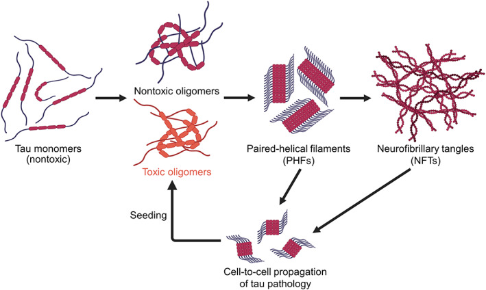
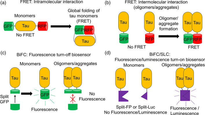
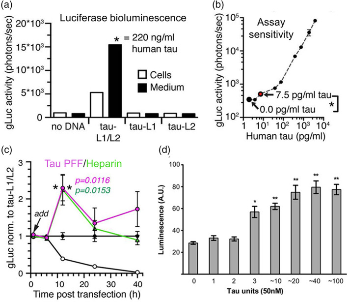
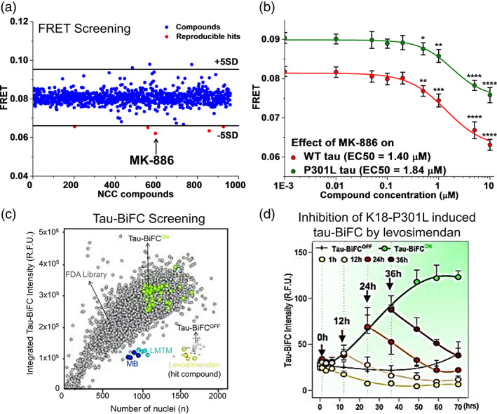
Tau is a microtubule binding protein which plays an important role in physiological functions but it is also involved in the pathogenesis of Alzheimer's disease and related tauopathies. While insoluble and β-sheet containing tau neurofibrillary tangles have been the histopathological hallmark of these diseases, recent studies suggest that soluble tau oligomers, which are formed prior to fibrils, are the primary toxic species. Substantial efforts have been made to generate tau oligomers using purified recombinant protein strategies to study oligomer conformations as well as their toxicity. However, no specific toxic tau species has been identified to date, potentially due to the lack of cellular environment. Hence, there is a need for cell-based models for direct monitoring of tau oligomerization and aggregation. This review will summarize the recent advances in the cellular biosensor technology, with a focus on fluorescence resonance energy transfer, bimolecular fluorescence complementation, and split luciferase complementation approaches, to monitor formation of tau oligomers and aggregates in living cells. We will discuss the applications of the cellular biosensors in examining the heterogeneous tau conformational ensembles and factors affecting tau self-assembly, as well as detecting cell-to-cell propagation of tau pathology. We will also compare the advantages and limitations of each type of tau biosensors, and highlight their translational applications in biomarker development and therapeutic discovery.

Keywords: Alzheimer's disease (AD); bimolecular fluorescence complementation (BiFC); cell‐based biosensor; conformational ensembles; fluorescence resonance energy transfer (FRET); high‐throughput screening (HTS); protein–protein interaction (PPI); split fluorescent protein complementation; split luciferase complementation (SLC); tau oligomerization.


Conflict of interest statement

The author declares no conflict of interest.

Figures

FIGURE 1
FIGURE 1
Tau fibrillogenesis cascade and cell‐to‐cell propagation of tau pathology in Alzheimer's disease. Misfolded tau species is capable of forming both nontoxic and toxic soluble tau oligomers spontaneously. The tau oligomers can proceed to form paired helical filaments (PHFs) and neurofibrillary tangles (NFTs) which are large insoluble aggregates with β‐sheet conformations. The fibrillar species can be secreted by host cells and transmitted to recipient cells which is capable of inducing further seeded oligomerization or aggregation, leading to cell‐to‐cell propagation of tau pathology. Schematics are created with BioRender.com
FIGURE 2
FIGURE 2
Schematic representation of tau biosensors based on fluorescence resonance energy transfer (FRET), bimolecular fluorescence complementation (BiFC), or split luciferase complementation (SLC) for monitoring intramolecular and intermolecular tau interactions in living cells. (a) Tau intramolecular FRET biosensor where FRET is observed when there is global folding of wild‐type (WT) monomeric tau. (b) Tau intermolecular FRET biosensors where FRET is observed when tau oligomers or aggregates are formed. WT tau is used for the formation of non‐β‐sheet soluble tau oligomers and tau repeat domain (tauRD) with P301S mutation or truncated tau is used for the formation of β‐sheet insoluble tau aggregates., , (c) BiFC tau fluorescence turn‐off biosensor where fluorescence is absent when there is tau oligomerization or aggregation., (d) BiFC/SLC tau fluorescence/luminescence turn‐on biosensor where fluorescence or luminescence is present when tau oligomers or aggregates are formed., , Tau oligomer is drawn as a dimer for illustration but it can be any species more than a dimer (≥2‐mers)
FIGURE 3
FIGURE 3
Cellular tau fluorescence resonance energy transfer (FRET) biosensors to examine tau oligomerization and aggregation. (a) Through acceptor photobleaching method, basal FRET is observed in both 0N4R wild‐type (WT) tau (T4) and Δ421‐441 truncated tau (T4C3) CFP/YFP FRET biosensors in the presence of kinase‐dead (Kd) glycogen synthase kinase 3 beta (GSK3β) (absence of GSK3β kinase activity). FRET efficiencies are increased with treatment of active GSK3β. (b) T4 FRET biosensors form soluble tau species or oligomers while T4C3 FRET biosensors form insoluble aggregates in the presence of active GSK3β. (c) Lifetime measurement of the WT 2N4R tau intermolecular GFP/RFP FRET biosensor shows a shorter lifetime in tau‐GFP/RFP (donor‐acceptor) sample as compared to tau‐GFP (donor) sample which exhibits efficient FRET. (d) Thioflavin‐S (ThS) staining of human embryonic kidney 293 (HEK293) cells expressing tau‐RFP (same total DNA concentration as the FRET biosensor) shows a positive signal with treatment of tau preformed fibrils (PFF) as a positive control. In the absence of PFF, there is no ThS signal, indicating formation of nonβ‐sheet soluble oligomers by the WT 2N4R tau FRET biosensor. (e) FRET measurement of tauRD CFP/YFP FRET biosensors with different tau variants expressed in HEK293 cells. PP refers to ΔK280/I227P/I308P mutations, ΔK refers to ΔK280, and LM refers to P301L/V337M mutations. (f) Formation of inclusions are observed in WT, ΔK and LM variants of the tauRD CFP/YFP FRET biosensors, as characterized by positive staining of X‐34 which is an amyloid‐specific dye. Permissions obtained from References , , and
FIGURE 4
FIGURE 4
Bimolecular fluorescence complementation (BiFC) biosensors to evaluate tau oligomerization and aggregation in cells and in a transgenic mouse model. (a) Fluorescence microscopy images of Venus‐based tau‐BiFC fluorescence turn‐on biosensor illustrating increased fluorescence complementation with treatment of phosphorylation inducers (okadiac acid at 30 nM and forskolin at 20 μM, scale bar = 200 μm). (b) Quantification of Venus‐based tau‐BiFC biosensor shows an increasing fluorescence signals with longer incubation time in cells treated both with and without phosphorylation inducers. (c) Schematic of human tauP301L‐BiFC transgenic mouse model which exhibits Venus fluorescence upon tau oligomerization and aggregation. Correlation plot of the immunofluorescence intensity profile between tau (green) and NeuN (red) shows the expression of tau in the cortex. (d) Representative images of fluorescence complementation in tauP301L‐BiFC mouse cortical regions at ages 3–12 months corresponding to different tau pathologies (scale bar = 50 μm). Permissions obtained from References and
FIGURE 5
FIGURE 5
Characterization of split luciferase complementation (SLC) biosensors and their ability to measure the minimum tau units to induce aggregation. (a) Split Gaussia luciferase (split‐gLuc) complementation biosensor illustrating increased complementation of luciferase bioluminescence both in human embryonic kidney 293 (HEK293) cells expressing the split gLuc plasmids (tau‐L1/L2) and in the culture medium where tau oligomers are released by the transfected cells. (d) Linear correlation between split‐gLuc activity and tau concentration indicating an assay sensitivity of 7.5 pg/ml tau‐L1/L2 equivalent to 0.16 nM full‐length tau as characterized by human total tau ELISA. (c) Treatment of tau preformed fibrils (PFF) and heparin to split‐gLuc biosensor accelerates oligomer formation in cell culture medium after 12 h with a subsequent decrease in the luciferase activity after 24 h. (d) Treatment of tauRD oligomers with a minimum of 3 units to HEK293 cells expressing RD‐Nluc/Cluc increases click beetle green luciferase signals. Permissions obtained from References and
FIGURE 6
FIGURE 6
Comparison of the sensitivity of the different cellular biosensors in detecting tau seeded aggregation. (a) Treatment of an increasing concentration of tauRD fibrils to human embryonic kidney 293 (HEK293) cells expressing tauRD‐Nluc/Cluc induces aggregation of the split luciferase complementation (SLC) biosensor as shown by an increase in the luciferase luminescence. (b) Treatment of an increasing concentration of tauRD fibrils to HEK293 cells expressing tauRD(ΔK)‐CFP/YFP induces aggregation of the fluorescence resonance energy transfer (FRET) biosensor. The ΔK indicates ΔK280 variant of tau. (c) Dose‐dependent increase in tau‐bimolecular fluorescence complementation (BiFC) fluorescence induced with tau K18‐P301L proteins in Venus‐based tau‐BiFC fluorescence turn‐on biosensor expressed in HEK293 cells. Permissions obtained from References , , and
FIGURE 7
FIGURE 7
Translational applications of fluorescence resonance energy transfer (FRET) and tau‐bimolecular fluorescence complementation (BiFC) biosensors in high‐throughput screening drug discovery. (a) High‐throughput screening of NIH Clinical Collection (NCC) library containing 727 compounds using wild‐type (WT) 2N4R tau intermolecular FRET biosensor expressed in human embryonic kidney 293 (HEK293) cells to obtain a small molecule inhibitor (MK‐886) of tau oligomerization. (b) MK‐886 reduces FRET in both 2N4R WT and P301L tau intermolecular biosensors with EC50 values of 1.40 and 1.84 μM, respectively. (c) High‐throughput screening of 1018 FDA approved compounds using Venus‐based tau‐BiFC fluorescence turn‐on biosensor expressed in HEK293 cells. Tau‐BiFCOFF (+), Tau‐BiFCON (green), FDA library (gray), methylene blue (MB, blue), LMTM (cyan), and levosimendan (hit compound, yellow) are indicated on the plot. (d) Treatment of levosimendan at different timings along the aggregation cascade of K18‐P301L induced tau aggregation in tau‐BiFC cells. Inhibitory effect of levosimendan is characterized by decreases in the tau‐BiFC responses. Permissions obtained from References and

References

    1. National Institute on Aging, NIH . Alzheimer's Disease Fact Sheet. 2021.
    1. Huang L‐K, Chao S‐P, Hu C‐J. Clinical trials of new drugs for Alzheimer disease. J Biomed Sci. 2020;27(1):18. - PMC - PubMed
    1. Cummings J, Lee G, Ritter A, Sabbagh M, Zhong K. Alzheimer's disease drug development pipeline: 2020. Alzheimer's Dement. 2020;6(1):e12050. - PMC - PubMed
    1. Ballatore C, Lee VMY, Trojanowski JQ. Tau‐mediated neurodegeneration in Alzheimer's disease and related disorders. Nat Rev Neurosci. 2007;8(9):663‐672. - PubMed
    1. Avila J, Lucas JJ, Pérez M, Hernández F. Role of tau protein in both physiological and pathological conditions. Physiol Rev. 2004;84(2):361‐384. - PubMed

LinkOut - more resources