. 2021 Jun 11;6(3):e10234.
doi: 10.1002/btm2.10234. eCollection 2021 Sep.

E3 ligase activity of Carboxyl terminus of Hsc70 interacting protein (CHIP) in Wharton's jelly derived mesenchymal stem cells improves their persistence under hyperglycemic stress and promotes the prophylactic effects against diabetic cardiac damages

Affiliations

E3 ligase activity of Carboxyl terminus of Hsc70 interacting protein (CHIP) in Wharton's jelly derived mesenchymal stem cells improves their persistence under hyperglycemic stress and promotes the prophylactic effects against diabetic cardiac damages

Ayaz Ali et al. Bioeng Transl Med. .

Abstract

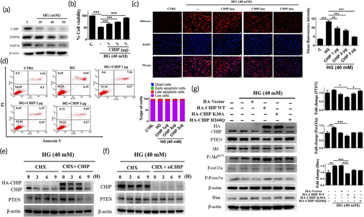
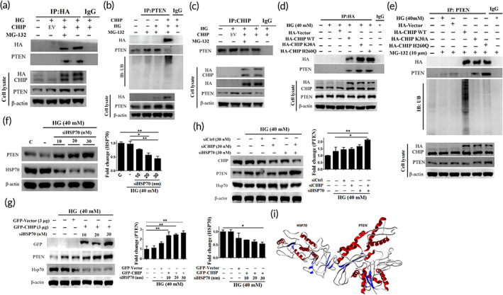
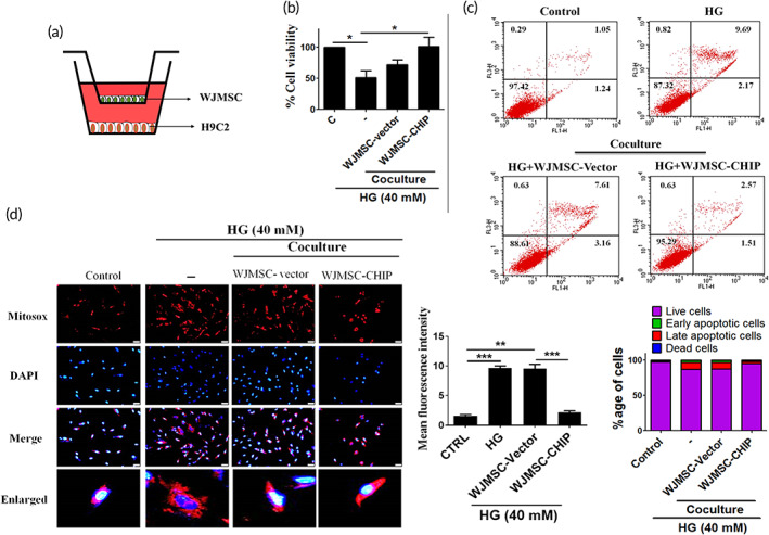
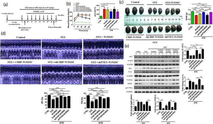
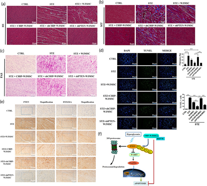
Recent studies indicate that umbilical cord stem cells are cytoprotective against several disorders. One critical limitation in using stem cells is reduction in their viability under stressful conditions, such as diabetes. However, the molecular intricacies responsible for diabetic conditions are not fully elucidated. In this study, we found that high glucose (HG) conditions induced loss of chaperone homeostasis, stabilized PTEN, triggered the downstream signaling cascade, and induced apoptosis and oxidative stress in Wharton's jelly derived mesenchymal stem cells (WJMSCs). Increased Carboxyl terminus of Hsc70 interacting protein (CHIP) expression promoted phosphatase and tensin homolog (PTEN) degradation via the ubiquitin-proteasome system and shortened its half-life during HG stress. Docking studies confirmed the interaction of CHIP with PTEN and FOXO3a with the Bim promoter region. Further, it was found that the chaperone system is involved in CHIP-mediated PTEN proteasomal degradation. CHIP depletion stabilizes PTEN whereas PTEN inhibition showed an inverse effect. CHIP overactivation suppressed the binding of FOXO3a with bim. Coculturing CHIP overexpressed WJMSCs suppressed HG-induced apoptosis and oxidative stress in embryo derived cardiac cell lines. CHIP overexpressing and PTEN silenced WJMSCs ameliorated diabetic effects in streptozotocin (STZ) induced diabetic rats and further improved their body weight and heart weight, and rescued from hyperglycemia-induced cardiac injury. Considering these, the current study suggests that CHIP confers resistance to apoptosis and acts as a potentiation factor in WJMSCs to provide protection from degenerative effects of diabetes.

Keywords: Wharton's jelly derived mesenchymal stem cells; apoptosis; carboxyl terminus of Hsc70 interacting protein; diabetes; phosphatase and tensin homolog.


Conflict of interest statement

The authors have no conflicts of interest to declare.

Figures

FIGURE 1
FIGURE 1
Effect of HG on PTEN‐mediated apoptosis and oxidative stress in WJMSCs. (a) WJMSCs seeded under varying concentration of HG for the indicated time points (24, 48, and 72 h) were harvested, and the cell viability was performed. (b, c) WJMSCs challenged with increasing concentrations of HG (30, 40, and 50 mM) for 24 h were incubated with the annexin V and PI and MitoSOX staining dye. The cell apoptosis and mitochondrial ROS were analyzed using flow cytometry and fluorescence microscopy. (d, e) WJMSCs were challenged with HG for 24 h, and the total cell lysate was harvested to analyze the expression of PTEN and downstream signaling cascade (AKT, p‐AKT, FOXO3a, p‐FOXO3a, and bim) via immunoblotting. (f) WJMSCs seeded in the presence of cycloheximide (CHX) were incubated with either MG‐132 (10 μM) or HG (40 mM) for the indicated time (0, 3, 6, 9 h) and thereafter immunoblotted with the anti‐PTEN antibody. β‐actin served as a loading control. The scale bar indicates 50 μm. Values shown are means ± SD. Quantification of the results are shown (n = 3). *p < 0.05, **p < 0.01, and ***p < 0.001 indicates the significant difference. HG, high glucose; PI, propidium iodide; PTEN, phosphatase and tensin homolog; ROS, reactive oxygen species; WJMSCs, Wharton's jelly derived mesenchymal stem cells
FIGURE 2
FIGURE 2
CHIP overexpressed WJMSCs attenuate hyperglycemia‐induced PTEN mediated apoptosis and oxidative stress. (a) WJMSCs were challenged with increasing concentrations of HG for 24 h, and subsequently the expression level of the chaperone system (HSP70, HSP90, and CHIP) was measured using Western blotting. (b) WJMSCs were transfected with varying increasing concentration of CHIP (1, 2, and 3 μg) followed by HG incubation for 24 h. The cell viability was detected. (C, D) WJMSCs were transfected with HA‐CHIP in the presence of HG (40 mM) for 24 h, and the mitochondrial ROS accumulation as well as apoptotic cell death were assessed using MitoSOX staining and flow cytometry. (e, f) WJMSCs were transfected with either HA‐CHIP (3 μg) or siCHIP (30 nM) for 24 h in the presence of CHX (50 μg/ml) for indicated time points were subjected to HG challenge for 24 h, and the protein expression was measured using Western blot analysis. (g) WJMSCs were transfected with pRK5‐HA‐vector (3 μg), pRK5‐HA‐CHIP (3 μg), pRK5‐HA‐K30A (3 μg), and pRK5‐HA‐H260Q (3 μg), followed by HG incubation for 24 h. Cell lysates were immunoblotted to analyze the expression of PTEN and the downstream signaling cascade. β‐actin act as a loading control. The scale bar indicates 100 μm. Values shown are means ± SD and quantification of the results shown as n = 3. *p < 0.05, **p < 0.01, and ***p < 0.001 indicates the significant difference. CHIP, carboxyl terminus of Hsc70 interacting protein; HG, high glucose; PTEN, phosphatase and tensin homolog; ROS, reactive oxygen species; WJMSCs, Wharton's jelly derived mesenchymal stem cells
FIGURE 3
FIGURE 3
CHIP regulates PTEN and its downstream signaling mediators under HG conditions. (a) Cells were transfected with increasing amounts of siCHIP (10, 20, 30 nM), and cell viability was determined after challenged with HG for 24 h. (b) WJMSCs incubated with varying increasing concentration of PTEN inhibitor (10, 25, 50 nM) were subjected to HG for 24 h, and the total cellular extract was analyzed using Western blot analysis. (c, d) Cells transfected with HA‐vector (3 μg), HA‐CHIP (3 μg), and shCHIP (3 μg) were challenged with HG for 24 h, and flow cytometry as well as MitoSOX staining was performed to analyze the apoptosis rate and mitochondrial ROS production. (e) WJMSCs were transfected with empty vector (EV) (3 μg), HA‐CHIP (3 μg), sicontrol (siCtrl) (30 nM), or siCHIP (30 nM) in the presence of HG for 24 h. Whole cell lysate was analyzed via immunoblotting. β‐actin served as a loading control. The scale bar indicates 100 μm. Values shown are mean ± SD. Quantification of the results are shown (n = 3). *p < 0.05, **p < 0.01, and ***p < 0.001 indicates the significant difference. CHIP, carboxyl terminus of Hsc70 interacting protein; HG, high glucose; PTEN, phosphatase and tensin homolog; ROS, reactive oxygen species; WJMSCs, Wharton's jelly derived mesenchymal stem cells
FIGURE 4
FIGURE 4
CHIP targets HG induced‐PTEN for ubiquitin‐mediated proteasomal degradation cooperated by HSP70 under HG conditions. (a–c) Cells transfected with HA‐vector or HA‐CHIP in the presence and absence of MG‐132 for 6 h were subjected to HG challenge for 24 h. Whole cell lysate was immunoprecipitated with the anti‐HA, anti‐CHIP, and anti‐PTEN antibody, and subsequently immunoblotted with the primary antibodies, including anti‐HA, anti‐PTEN, and anti‐ubiquitin. (d, e) Cells transfected with HA‐vector, HA‐CHIP, and CHIP mutants (K30A, an H260Q) were treated with or without MG‐132 for 6 h in the presence of HG for 24 h. Whole cell lysate was immunoprecipitated with the anti‐HA and anti‐PTEN antibody followed by immunoblotting with the anti‐HA, anti‐PTEN, and anti‐ubiquitin antibody. (f) Cells were transfected with increasing concentrations of siHSP70 (10, 20, 30 nM) after challenged with HG for 24 h, and the expression level of PTEN and HSP70 was measured employing Western blot analysis. (g) Following cotransfection of GFP‐vector or GFP‐CHIP with increasing concentration of siHSP70 in WJMSCs were challenged with HG for 24 h, and the protein expression was measured via immunoblotting. (h) WJMSCs transfected with sicontrol, CHIP siRNA, or siHSP70 were subjected to HG challenge for 24 h, and the total cell extract was immunoblotted with CHIP, PTEN, and HSP70. β‐actin served as a loading control. (i) Docking studies demonstrating the molecular interaction of HSP70 with PTEN forming a heteromer complex (HSP70 and PTEN shown in quaternary structure with helices and sheets in complex). Values shown are mean ± SD. Quantification of the results are shown (n = 3). *p < 0.05, **p < 0.01, and ***p < 0.001 indicates the significant difference. CHIP, carboxyl terminus of Hsc70 interacting protein; HG, high glucose; PTEN, phosphatase and tensin homolog; WJMSCs, Wharton's jelly derived mesenchymal stem cells
FIGURE 5
FIGURE 5
CHIP regulates the binding of FOXO3a with the Bim promoter region. (a) WJMSCs transfected with increasing amounts of siFOXO3a were challenged with HG stress, and the expression levels of FOXO3a and Bim were assessed by immunoblotting. (b) Cells transfected with shcontrol or siFOXO3a in the presence and absence of LY294002 (PI3K inhibitor) were subjected to HG for 24 h, and the activation of FOXO3a, p‐FOXO3a, and Bim was analyzed using Western blot assay. (c) WJMSCs transfected with HA‐vector, HA‐CHIP, shcontrol, shCHIP, or siFOXO3a were incubated with HG for 24 h followed by immunoblotting to analyze the HA, FoxO3a, and Bim levels. (d) WJMSCs were transfected with varying concentration of siCHIP (10, 20, 30 nM) in the presence of HG for 24 h. Thereafter, the extracted cell lysates using cytoplasmic and nuclear fractionation kit were immunoblotted. (e) WJMSCs transfected with HA‐CHIP or shCHIP plasmids were challenged with HG for 24 h and ChIP assay was performed to evaluate the FOXO3a interaction with the Bim promoter region. (f) Prediction of Bim promoter region using NCBI database (brown color indicates Bim promoter region) and converted to three‐dimensional structure for molecular interaction with FOXO3a. (g) Docking studies illustrating the molecular interaction between FOXO3a and the Bim promoter region (Bim region is shown in green color and FOXO3a shown in red color). *p < 0.05, **p < 0.01, and ***p < 0.001 shows the significance. CHIP, carboxyl terminus of Hsc70 interacting protein; HG, high glucose; WJMSCs, Wharton's jelly derived mesenchymal stem cells
FIGURE 6
FIGURE 6
Coculturing CHIP overexpressed WJMSCs with cardiac cells rescued HG‐induced cardiac apoptosis and oxidative stress. (a) Schematic representation of in vitro coculturing WJMSCs with H9c2 cardiomyoblasts. (b) WJMSCs transfected with HA‐vector or HA‐CHIP in the presence of HG were cocultured with cardiomyoblasts followed by incubation with MTT reagent to assess the cell viability. (c, d) Embryo derived cardiac cells were cocultured with WJMSCs alone, WJMSCs transfected with HA‐vector, or HA‐CHIP in the presence of HG stress for 24 h. Flow cytometry and MitoSOX staining were performed to estimate the apoptotic cell death and mitochondrial oxidative stress generation. The scale bar indicates 50 μm. β‐actin acts as a loading control. Values shown are mean ± SD. Quantification of the results are shown (n = 3). *p < 0.05, **p < 0.01, and ***p < 0.001 represents the significance. CHIP, carboxyl terminus of Hsc70 interacting protein; HG, high glucose; WJMSCs, Wharton's jelly derived mesenchymal stem cells
FIGURE 7
FIGURE 7
CHIP overexpressed WJMSCs rescued hyperglycemic effects under diabetic conditions. (a) Schematic illustration of STZ‐induced diabetes, and WJMSCs administration expressing different plasmids, including GFP‐CHIP, shCHIP, and shPTEN. (b) The oral glucose tolerance test (OGTT) was performed after 6 weeks treatment for the indicated time points (0, 30, 60, 90, and 120 min) in various experimental groups, including control, STZ‐induced diabetes (STZ), STZ‐induced diabetes administered with WJMSCs alone (STZ + WJMSCs), STZ‐induced diabetes injected with CHIP overexpressed WJMSCs (STZ + CHIP‐WJMSCs), STZ‐induced diabetes transplanted with CHIP knockdown WJMSCs (STZ + shCHIP‐WJMSCs), and STZ‐induced diabetic rats infused with PTEN knockdown WJMSCs (STZ + shPTEN‐WJMSCs) after the rats were fasted for 14 h. (c) Morphological assessment of cardiac tissues in different experimental groups. (d) Echocardiographic evaluation of cardiac function in different experimental groups (control, STZ, STZ + WJMSCs, STZ + CHIP‐WJMSCs, STZ + shCHIP‐WJMSCs, and STZ + shPTEN‐WJMSCs). (e) Total cell lysate from the left ventricle was quantified and measured using Western blot. Protein expression levels of the apoptosis (p‐AKT, Bax, and Cyt‐c) and oxidative stress markers (catalase, SOD2, and gp91PHOX) were assessed. GAPDH act as a loading control. Values shown are mean ± SD. Quantification of the results are shown (n = 3). *p < 0.05, **p < 0.01, and ***p < 0.001 shows the significance. CHIP, carboxyl terminus of Hsc70 interacting protein; GAPDH, Glyceraldehyde‐3‐phosphate dehydrogenase; PTEN, phosphatase and tensin homolog; STZ, streptozotocin; WJMSCs, Wharton's jelly derived mesenchymal stem cells
FIGURE 8
FIGURE 8
CHIP overexpressing WJMSCs ameliorated hyperglycemia‐induced cardiac injuries in diabetic animals. (a–c) Hematoxylin‐eosin (HE), Masson's trichrome (MT), and Periodic acid‐Schiff staining (PAS) were performed to evaluate cardiac morphology, fibrosis, and glycogen accumulation in different experimental groups. (d) Cardiomyocyte apoptosis was assessed using the TUNEL assay. (e) Immunohistochemistry assay (IHC) was performed to evaluate cardiac expression of PTEN and FOXO3a in different experimental groups. (f) Schematic illustration of CHIP‐mediated PTEN degradation under hyperglycemic conditions showed resistance to apoptosis. The scale bar indicates 50 μm. Values shown are mean ± SD. Quantification of the results are shown (n = 3). *p < 0.05, **p < 0.01, and ***p < 0.001 indicates the significant difference. CHIP, carboxyl terminus of Hsc70 interacting protein; PTEN, phosphatase and tensin homolog; TUNEL, Terminal deoxynucleotidyl transferase dUTP nick end labeling; WJMSCs, Wharton's jelly derived mesenchymal stem cells

References

    1. Einarson TR, Acs A, Ludwig C. Prevalence of cardiovascular disease in type 2 diabetes: a systematic literature review of scientific evidence from across the world in 2007–2017. Cardiovasc Diabetol. 2018;17(1):83. - PMC - PubMed
    1. Abdul‐Ghani M, DeFronzo RA, Del Prato S, et al. Cardiovascular disease and type 2 diabetes: has the dawn of a new era arrived? Diabetes Care. 2017;40:813‐820. - PMC - PubMed
    1. Filardi T, Ghinassi B. Cardiomyopathy associated with diabetes: the central role of the cardiomyocyte. Int J Mol Sci. 2019;20(13):3299. - PMC - PubMed
    1. Lyons TJ, Basu A. Biomarkers in diabetes: hemoglobin A1c, vascular and tissue markers. Transl Res. 2012;159(4):303‐312. - PMC - PubMed
    1. Yang YC, Tsai CY, Chen CL, et al. Pkcδ activation is involved in ROS‐mediated mitochondrial dysfunction and apoptosis in cardiomyocytes exposed to advanced glycation end products (ages). Aging Dis. 2018;9(4):647‐663. - PMC - PubMed