. 2020 May;1(3):99-113.
doi: 10.1007/s42764-020-00009-8. Epub 2020 Mar 19.

Mechanisms of UV-induced mutations and skin cancer

Affiliations

Mechanisms of UV-induced mutations and skin cancer

Gerd P Pfeifer. Genome Instab Dis. 2020 May.

Abstract

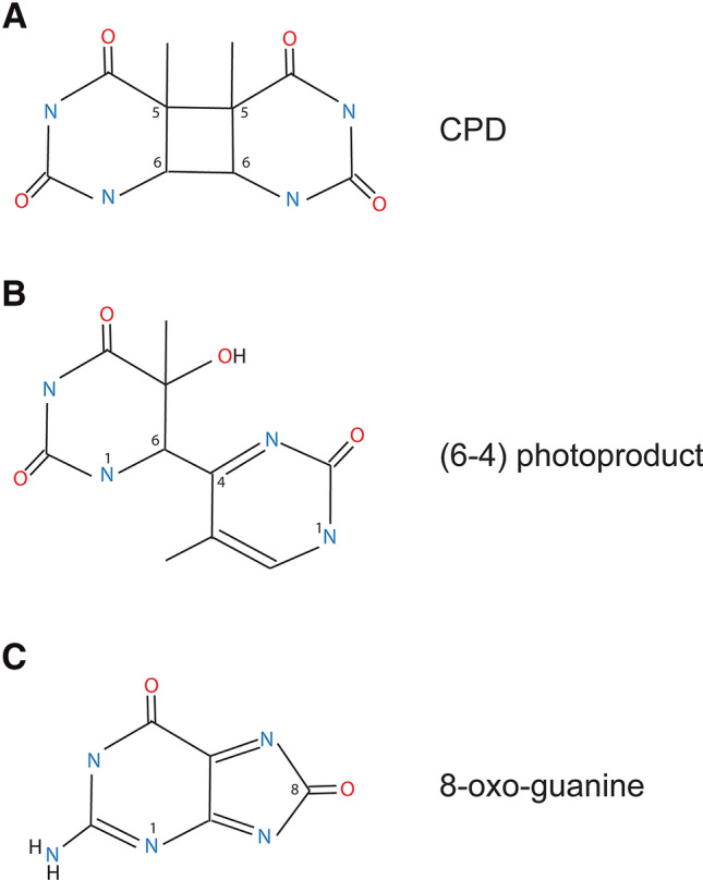
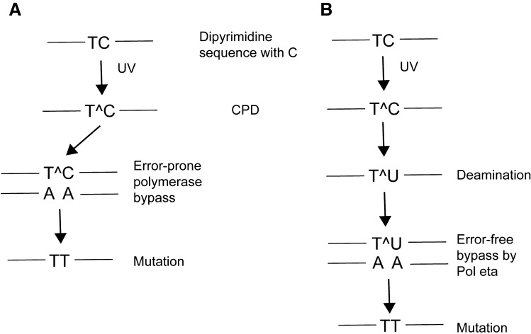
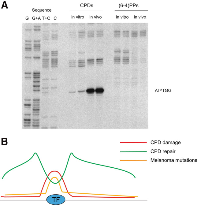
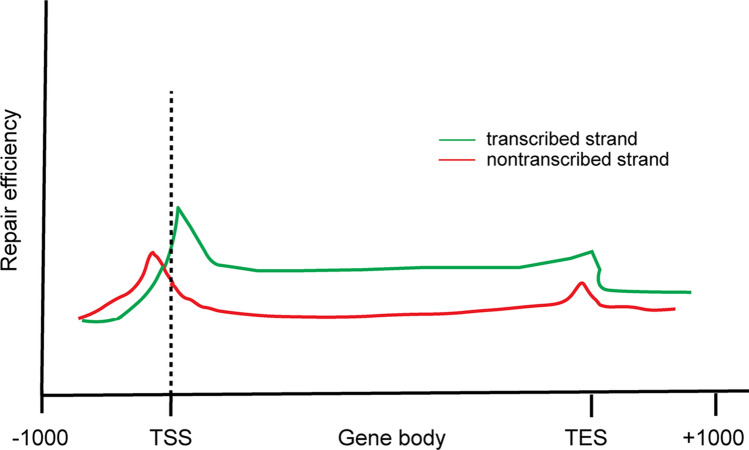
Ultraviolet (UV) irradiation causes various types of DNA damage, which leads to specific mutations and the emergence of skin cancer in humans, often decades after initial exposure. Different UV wavelengths cause the formation of prominent UV-induced DNA lesions. Most of these lesions are removed by the nucleotide excision repair pathway, which is defective in rare genetic skin disorders referred to as xeroderma pigmentosum. A major role in inducing sunlight-dependent skin cancer mutations is assigned to the cyclobutane pyrimidine dimers (CPDs). In this review, we discuss the mechanisms of UV damage induction, the genomic distribution of this damage, relevant DNA repair mechanisms, the proposed mechanisms of how UV-induced CPDs bring about DNA replication-dependent mutagenicity in mammalian cells, and the strong signature of UV damage and mutagenesis found in skin cancer genomes.

Keywords: (6–4) photoproduct; DNA repair; UV; Ultraviolet; cyclobutane pyrimidine dimer; melanoma; mutations; skin cancer.


Figures

Fig. 1
Fig. 1
The major DNA damage products induced by solar UVB irradiation. a Cyclobutane pyrimidine dimer (CPD) at TT sequences. b Pyrimidine (6–4) pyrimidone photoproduct [(6–4)PP] at TT sequences. c 8-Oxoguanine
Fig. 2
Fig. 2
Pathways leading to UV mutagenesis at CPDs containing cytosine. a In the error-prone DNA synthesis pathway, a DNA polymerase bypasses the CPD by insertion of an incorrect adenine base across the cytosine leading to C to T mutations. b In the deamination-bypass pathway, the CPD that has formed at dipyrimidines containing cytosine undergoes hydrolytic deamination to uracil. DNA synthesis past uracil-containing dimers by DNA polymerase eta occurs in an error-free manner by incorporation of adenine across uracil. However, the mutation is fixed due to the deamination event. Note that a similar pathway may operate at CPDs containing 5-methylcytosine. In that case, deamination leads to the formation of thymine within the dimers followed by error-free Pol eta bypass of the lesion. UV-induced CC to TT mutations may arise from double cytosine or 5-methylcytosine deamination events
Fig. 3
Fig. 3
UV damage at transcription factor binding sites and melanoma mutations. a A region 100–150 base pairs upstream of the transcription start site of the PCNA gene acquires high levels of CPDs after UV irradiation of HeLa cells at a specific position containing a consensus binding site of the NFY transcription factor (5′ATTGG). CPDs and (6–4)PPs were mapped separately in this region (Tornaletti and Pfeifer 1995). Note that these CPDs at 5′TT sequences would not be mutagenic. b Schematic illustration of high levels of UV damage (CPDs) at transcription factor (TF) binding sites genome-wide, generally enhanced repair in open chromatin regions surrounding the TF but diminished repair at the TF binding site itself, and the resulting mutation hotspots targeted to such binding sites in melanoma skin tumor genomes (Sabarinathan et al. ; Elliott et al. 2018)
Fig. 4
Fig. 4
Genome-wide maps of nucleotide excision repair of CPDs. The XR-seq method was used to obtain genome-wide single nucleotide resolution maps of repair of CPDs. The figure shows a schematic illustration of the data obtained by Hu et al. (2015) for excision repair of CPDs in normal human fibroblasts. The graphs shown are average profiles for all UCSC reference genes. Note the more extensive repair of the transcribed DNA strand relative to the nontranscribed strand, the fast repair near the transcription start sites (TSS) and a slightly enhanced repair near the transcription end sites (TES). The faster repair of the nontranscribed strand upstream of the TSS is thought to be due to divergent transcription emanating from the promoters, which is a common feature of mammalian cells

References

    1. Adar S, Hu J, Lieb JD, Sancar A. Genome-wide kinetics of DNA excision repair in relation to chromatin state and mutagenesis. Proceedings of the National Academy of Sciences of the United States of America. 2016;113(15):E2124–2133. doi: 10.1073/pnas.1603388113. - DOI - PMC - PubMed
    1. Akagi JI, et al. Effect of sequence context on Polzeta-dependent error-prone extension past (6–4) photoproducts. DNA Repair (Amsterdam) 2019;87:102771. doi: 10.1016/j.dnarep.2019.102771. - DOI - PubMed
    1. Albert A, Knoll MA, Conti JA, Zbar RIS. Non-melanoma skin cancers in the older patient. Current Oncology Reports. 2019;21(9):79. doi: 10.1007/s11912-019-0828-9. - DOI - PubMed
    1. Araujo SJ, Kuraoka I. Nucleotide excision repair genes shaping embryonic development. Open Biology. 2019;9(10):190166. doi: 10.1098/rsob.190166. - DOI - PMC - PubMed
    1. Autier P, et al. Cutaneous malignant melanoma and exposure to sunlamps or sunbeds: An EORTC multicenter case–control study in Belgium, France and Germany. EORTC Melanoma Cooperative Group. International Journal of Cancer. 1994;58:809–813. doi: 10.1002/ijc.2910580610. - DOI - PubMed

LinkOut - more resources