Review
. 2021 Sep 23:14:26317745211045769.
doi: 10.1177/26317745211045769. eCollection 2021 Jan-Dec.

Management of pancreatic cysts and guidelines: what the gastroenterologist needs to know

Affiliations
Review

Management of pancreatic cysts and guidelines: what the gastroenterologist needs to know

Ross C D Buerlein et al. Ther Adv Gastrointest Endosc. .

Abstract

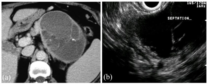
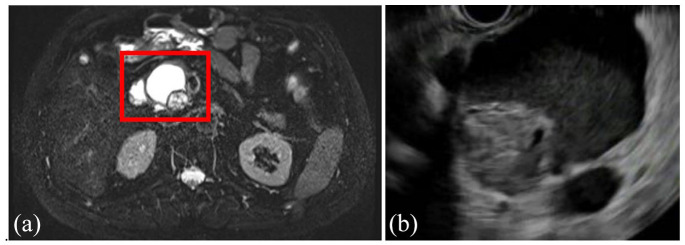
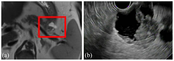
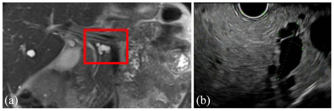
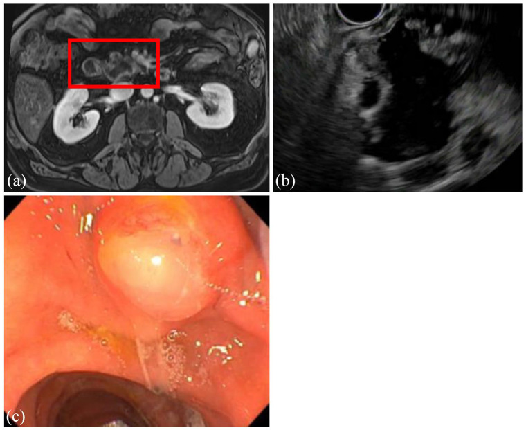
The prevalence of pancreatic cysts has increased significantly over the last decade, partly secondary to increased quality and frequency of cross-sectional imaging. While the majority never progress to cancer, a small number will and need to be followed. The management of pancreatic cysts can be both confusing and intimidating due to the multiple guidelines with varying recommendations. Despite the differences in the specifics of the guidelines, they all agree on several high-risk features that should get the attention of any clinician when assessing a pancreatic cyst: presence of a mural nodule or solid component, dilation of the main pancreatic duct (or presence of main duct intraductal papillary mucinous neoplasm), pancreatic cyst size ⩾3-4 cm, or positive cytology on pancreatic cyst fluid aspiration. Other important criteria to consider include rapid cyst growth (⩾5 mm/year), elevated serum carbohydrate antigen 19-9 levels, new-onset diabetes mellitus, or acute pancreatitis thought to be related to the cystic lesion.

Keywords: mucinous cystic neoplasms; pancreatic cyst; pancreatic cyst guidelines; pancreatic cystic neoplasms; pancreatic cystic tumors; pancreatic neoplasia.


Conflict of interest statement

Conflict of interest statement: The authors declared the following potential conflicts of interest with respect to the research, authorship, and/or publication of this article: Vanessa M Shami is consultant for both Interpace Diagnostics and Olympus America. Ross CD Buerlein has no conflicts of interest.

Figures

Figure 1.
Figure 1.
Demonstration of a positive ‘string sign’ where a drop of aspirated pancreatic cystic fluid is placed between two glass slides, and as the two glass slides are slowly pulled apart, there is a string of mucous >3.5 mm in length. This is consistent with a mucinous pancreatic cyst.
Figure 2.
Figure 2.
Coronal view of MRI (a) and EUS (b) showing a 4.2-cm serous cystic neoplasm with classic honeycombing and microcystic pattern in the uncinated process of the pancreas of a 57-year-old woman, which was discovered incidentally during the workup for a ventral hernia. She was asymptomatic, and no additional surveillance was recommended.
Figure 3.
Figure 3.
Axial view of CT (a) and EUS (b) of a 5.2-cm solid pseudopapillary neoplasm in a 43-year-old woman who presented with abdominal pain. The lesion is well demarcated with mixed solid and cystic features.
Figure 4.
Figure 4.
Coronal view of MRI (a) and EUS (b) of a cystic pancreatic neuroendocrine tumor found in a 55-year-old woman during the workup for abdominal pain. The lesion is well circumscribed and solitary.
Figure 5.
Figure 5.
Axial view of CT (a) and EUS (b) of a mucinous cystic neoplasm found incidentally in the tail of the pancreas of a 53-year-old woman. Several thin septations can be seen on both CT and EUS. The patient underwent lateral pancreatectomy which confirmed the diagnosis, and no malignancy was present.
Figure 6.
Figure 6.
Axial view of MRI (a) and EUS (b) showing a 4.7-cm BD-IPMN in the head of the pancreas with an associated mural nodule in a 62-year-old man who was incidentally found to have a pancreatic cystic lesion on imaging obtained as part of the workup for COPD. EUS-guided fine needle aspiration was consistent with poorly differentiated carcinoma. He subsequently underwent pancreatoduodenectomy, with pathology showing the same.
Figure 7.
Figure 7.
Axial view of MRI (a) and EUS (b) of a 3-cm branch duct IPMN in the tail of the pancreas with a solid component in a 79-year-old patient. The patient underwent EUS-guided fine needle aspiration of the solid component with cytology revealing adenocarcinoma. The patient then underwent lateral pancreatectomy with splenectomy and regional lymphadenectomy. Pathology revealed an invasive mucinous adenocarcinoma, and all lymph nodes were negative.
Figure 8.
Figure 8.
Coronal view of MRI (a) and EUS (b) showing a 2.3-cm branch duct IPMN in the body of the pancreas of a 69-year-old man with no worrisome or high-risk features.
Figure 9.
Figure 9.
Axial view of MRI (a) and EUS (b) of a main duct intraductal papillary mucinous neoplasm (MD-IPMN) identified in a 66-year-old man. The main pancreatic duct is dilated to 16.4 mm extending from the head to the genu of the pancreas. Endoscopic view (c) of the major papilla showing spontaneous extrusion of mucous (‘Mucorrhea’), which is pathognomonic for an MD-IPMN. This patient underwent a pancreatoduodenectomy with pathology revealing focally invasive carcinoma arising from an IPMN with high-grade dysplasia.
Figure 10.
Figure 10.
Timeline of major guideline publications related to the management of pancreatic cystic neoplasms. AGA, American Gastroenterological Association; ACG, American College of Gastroenterology.
Figure 11.
Figure 11.
Algorithm to approach of pancreatic cysts. *High-risk features: presence of mural nodule or solid component, dilation of main pancreatic duct (⩾5 mm), cyst size ⩾3–4 cm, and positive cytology on fluid aspiration.

References

    1. Ferrone CR, Correa-Gallego C, Warshaw AL, et al.. Current trends in pancreatic cystic neoplasms. Arch Surg 2009; 144: 448–454. - PMC - PubMed
    1. Kromrey ML, Bülow R, Hübner J, et al.. Prospective study on the incidence, prevalence and 5-year pancreatic-related mortality of pancreatic cysts in a population-based study. Gut 2018; 67: 138–145. - PubMed
    1. Stark A, Donahue TR, Reber HA, et al.. Pancreatic cyst disease: a review. JAMA 2016; 315: 1882–1893. - PubMed
    1. Tada M, Kawabe T, Arizumi M, et al.. Pancreatic cancer in patients with pancreatic cystic lesions: a prospective study in 197 patients. Clin Gastroenterol Hepatol 2006; 4: 1265–1270. - PubMed
    1. Matsubara S, Tada M, Akahane M, et al.. Incidental pancreatic cysts found by magnetic resonance imaging and their relationship with pancreatic cancer. Pancreas 2012; 41: 1241–1246. - PubMed

LinkOut - more resources