

Sex differences in a murine model of Cerebral Amyloid Angiopathy
- PMID: 34589766
- PMCID: PMC8474688
- DOI: 10.1016/j.bbih.2021.100260
Sex differences in a murine model of Cerebral Amyloid Angiopathy
Abstract
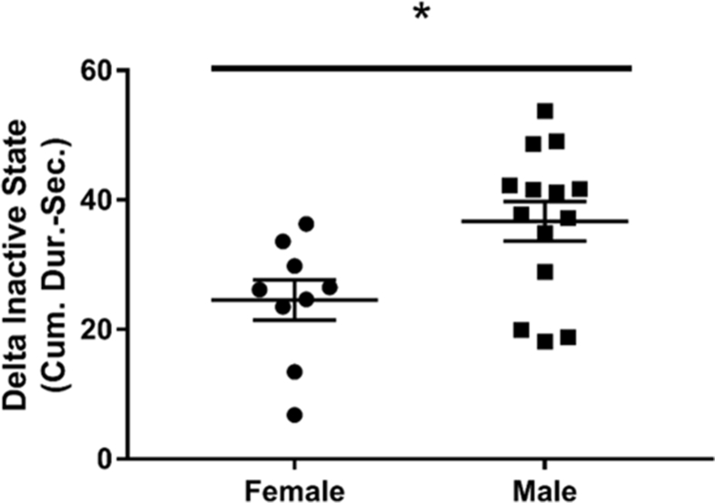
Cerebral amyloid angiopathy (CAA) is one of the common causes of lobar intracerebral hemorrhage and vascular cognitive impairment (VCI) in the aging population. Increased amyloid plaque deposition within cerebral blood vessels, specifically the smooth muscle layer, is linked to increased cerebral microbleeds (CMBs) and impaired cognition in CAA. Studies in Alzheimer's disease (AD) have shown that amyloid plaque pathology is more prevalent in the brains of elderly women (2/3rd of the dementia population) compared with men, however, there is a paucity of studies on sex differences in CAA. The objective of this study was to discern the sexual dichotomies in CAA. We utilized male and female Tg-SwDI mice (mouse model of CAA) at 12-14 months of age for this study. We evaluated sex differences in CMBs, cognitive function and inflammation. Cognition was assessed using Y-maze (spatial working memory) and Fear Conditioning (contextual memory). CMBs were quantified by ex vivo brain MRI scans. Inflammatory cytokines in brain were quantified using ELISA. Our results demonstrated that aging Tg-SwDI female mice had a significantly higher burden of CMBs on MRI as compared to males. Interestingly, these aging Tg-SwDI female mice also had significantly impaired spatial and contextual memory on Y maze and Fear Conditioning respectively. Furthermore, female mice had significantly lower circulating inflammatory cytokines, IL-1α, IL-2, IL-9, and IFN-γ, as compared to males. Our results demonstrate that aging female Tg-SwDI mice are more cognitively impaired and have higher number of CMBs, as compared to males at 12-14 months of age. This may be secondary to reduced levels of neural repair cytokines (IL-1α, IL-2, IL-9 and IFN-γ) involved in sex specific inflammatory signaling in CAA.
Keywords: Cerebral amyloid angiopathy; Cerebral microbleeds; Cognition; MRI.
© 2021 The Authors.
Conflict of interest statement
The authors declare that they have no known competing financial interests or personal relationships that could have appeared to influence the work reported in this paper.
Figures


Similar articles
-
Long-term voluntary wheel running does not alter vascular amyloid burden but reduces neuroinflammation in the Tg-SwDI mouse model of cerebral amyloid angiopathy.J Neuroinflammation. 2019 Jul 11;16(1):144. doi: 10.1186/s12974-019-1534-0. J Neuroinflammation. 2019. PMID: 31296239 Free PMC article.
-
Taxifolin inhibits amyloid-β oligomer formation and fully restores vascular integrity and memory in cerebral amyloid angiopathy.Acta Neuropathol Commun. 2017 Apr 4;5(1):26. doi: 10.1186/s40478-017-0429-5. Acta Neuropathol Commun. 2017. PMID: 28376923 Free PMC article.
-
Cerebral Amyloid Angiopathy Burden and Cerebral Microbleeds: Pathological Evidence for Distinct Phenotypes.J Alzheimers Dis. 2021;81(1):113-122. doi: 10.3233/JAD-201536. J Alzheimers Dis. 2021. PMID: 33720897 Free PMC article.
-
Cerebral amyloid angiopathy: major contributor or decorative response to Alzheimer's disease pathogenesis.Neurobiol Aging. 2004 May-Jun;25(5):599-602; discussion 603-4. doi: 10.1016/j.neurobiolaging.2003.12.019. Neurobiol Aging. 2004. PMID: 15172735 Review.
-
Prevalence of cerebral amyloid angiopathy: A systematic review and meta-analysis.Alzheimers Dement. 2022 Jan;18(1):10-28. doi: 10.1002/alz.12366. Epub 2021 May 31. Alzheimers Dement. 2022. PMID: 34057813 Free PMC article.
Cited by
-
Chronic Treatment of a Mouse Model of Cerebral Amyloid Angiopathy and Brain AT1 Receptor Expression.bioRxiv [Preprint]. 2025 May 21:2025.05.16.654535. doi: 10.1101/2025.05.16.654535. bioRxiv. 2025. PMID: 40475476 Free PMC article. Preprint.
-
"Let's talk about sex, inflammaging, and cognition, baby": A meta-analysis and meta-regression of 106 case-control studies on mild cognitive impairment and Alzheimer's disease.Brain Behav Immun Health. 2024 Jul 20;40:100819. doi: 10.1016/j.bbih.2024.100819. eCollection 2024 Oct. Brain Behav Immun Health. 2024. PMID: 39161876 Free PMC article. Review.
-
Neutrophils and Neutrophil Extracellular Traps Cause Vascular Occlusion and Delayed Cerebral Ischemia After Subarachnoid Hemorrhage in Mice.Arterioscler Thromb Vasc Biol. 2024 Mar;44(3):635-652. doi: 10.1161/ATVBAHA.123.320224. Epub 2024 Feb 1. Arterioscler Thromb Vasc Biol. 2024. PMID: 38299355 Free PMC article.
-
Sex differences in the progression of cerebral microbleeds in patients with concomitant cerebral small vessel disease.Front Neurol. 2022 Dec 20;13:1054624. doi: 10.3389/fneur.2022.1054624. eCollection 2022. Front Neurol. 2022. PMID: 36619919 Free PMC article.
-
Sex differences in age-associated neurological diseases-A roadmap for reliable and high-yield research.Sci Adv. 2025 Mar 7;11(10):eadt9243. doi: 10.1126/sciadv.adt9243. Epub 2025 Mar 5. Sci Adv. 2025. PMID: 40043111 Free PMC article. Review.
References
-
- Alves S., Churlaud G., Audrain M., Michaelsen-Preusse K., Fol R., Souchet B. Interleukin-2 improves amyloid pathology, synaptic failure and memory in Alzheimer’s disease mice. Brain. 2017;140(3):826–842. - PubMed
-
- Auriel E., Greenberg S.M. The pathophysiology and clinical presentation of cerebral amyloid angiopathy. Curr. Atherosclerosis Rep. 2012;14(4):343–350. - PubMed
-
- Barnes L.L., Wilson R.S., Bienias J.L., Schneider J.A., Evans D.A., Bennett D.A. Sex differences in the clinical manifestations of Alzheimer disease pathology. Arch. Gen. Psychiatr. 2005;62(6):685–691. - PubMed
-
- Baron R., Nemirovsky A., Harpaz I., Cohen H., Owens T., Monsonego A. IFN-gamma enhances neurogenesis in wild-type mice and in a mouse model of Alzheimer’s disease. Faseb. J. 2008;22(8):2843–2852. - PubMed
Grants and funding
LinkOut - more resources
Full Text Sources
Miscellaneous
