Review
. 2020 Mar 18:4:100060.
doi: 10.1016/j.bbih.2020.100060. eCollection 2020 Apr.

Lipopolysaccharide animal models of Parkinson's disease: Recent progress and relevance to clinical disease

Affiliations
Review

Lipopolysaccharide animal models of Parkinson's disease: Recent progress and relevance to clinical disease

Isaac Deng et al. Brain Behav Immun Health. .

Abstract

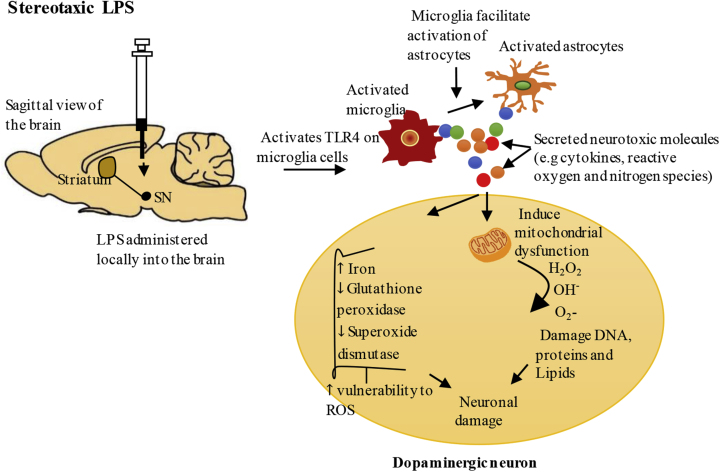
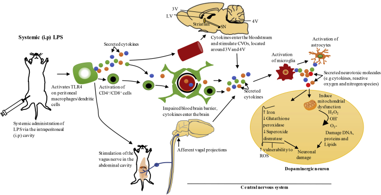
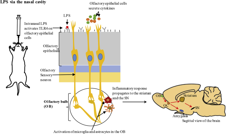
Parkinson's disease (PD) is one of the most common neurodegenerative movement disorders which is characterised neuropathologically by progressive loss of dopaminergic neurons in the substantia nigra pars compacta (SNpc) and the presence of Lewy bodies (made predominately of α-synuclein) in the surviving neurons. Animal models of PD have improved our understanding of the disease and have played a critical role in the development of neuroprotective agents. Neuroinflammation has been strongly implicated in the pathogenesis of PD, and recent studies have used lipopolysaccharide (LPS), a component of gram-negative bacteria and a potent activator of microglia cells, to mimic the inflammatory events in clinical PD. Modulating the inflammatory response could ameliorate PD associated complications and thus, it is essential to understand the extent to which LPS models reflect human PD. This review will outline the routes of administration of LPS such as stereotaxic, systemic and intranasal, their ability to recapitulate neuropathological markers of PD, and mechanisms of LPS induced toxicity. We will also discuss the ability of the models to replicate motor symptoms and non-motor symptoms of PD such as gastrointestinal dysfunction, olfactory dysfunction, anxiety, depression and cognitive dysfunction.

Keywords: Animal models; Lipopolysaccharide; Motor symptoms; Non-motor symptoms; Parkinson’s disease.


Conflict of interest statement

The authors declare that they have no known competing financial interests or personal relationships that could have appeared to influence the work reported in this paper.

Figures

Fig. 1
Fig. 1
Stereotaxicadministration ofLPS – the proposed mechanisms leading todopaminergicneuron degeneration. Stereotaxic administration of LPS (delivered directly into the local brain environment) can activate Toll-like receptor 4 (TLR4) on microglia cells resulting in the production of inflammatory cytokines (e.g.TNF-α, IL-1β, IL-6, IFN-γ) and free radicals (e.g. NO and O2-) in the brain (Liu and Bing, 2011). Activated microglia facilitate the activation of astrocytes which augment the inflammatory response through additional production of inflammatory cytokines, and reactive oxygen and nitrogen species. Excessive production of these neurotoxic molecules could impair mitochondrial function, resulting in impaired energy metabolism and additional production of reactive oxygen species (ROS), and can reduce antioxidants such as glutathione peroxidase and superoxide dismutase in the brain (Hauser and Hastings, 2013; Park et al., 2018; Villacé et al., 2017). Stereotaxic LPS also increases iron levels in the substantia nigra (SN), which could catalyse the Fenton reaction of superoxide (O2-) and hydrogen peroxide (H2O2) to yield hydroxyl radicals (OH) which are toxic to the neurons (Hauser and Hastings, 2013).
Fig. 2
Fig. 2
Systemic administration of LPS via intraperitoneal cavity-the proposed pathways of propagation of systemic inflammation to the brain. Systemic administration of LPS via intraperitoneal (i.p) cavity activates Toll-like receptor 4 (TLR4) on peritoneal macrophages/dendritic cells which results in local secretion of inflammatory cytokines such as IL-1, TNF-α and IL-6 (Block et al., 2007; Konsman et al., 1999). Systemic inflammation could be communicated to the brain through several mechanisms. Firstly, IL-1 produced by peritoneal macrophages/dendritic cells at the site of injection in response to systemic LPS, can activate IL-1 receptor on the vagus nerve that innervates the abdominal cavity. This nerve terminates in the nucleus of solitary tract located in the brainstem, which projects to other brain regions that mediate acute sickness response (Konsman et al., 2002; Vitkovic et al., 2000). Secondly, the blood brain barrier (BBB) could lose its integrity under pathological conditions, resulting in the entry of peripheral immune cells such as CD4+/CD8+ T-lymphocytes and inflammatory cytokines into the brain, and these molecules can mediate neuroinflammation through activation of microglia and astrocytes (Hoban et al., 2013). Furthermore, systemic cytokines can communicate with circumventricular organs (CVOs) in the CNS, which are a group of structures located around the third and fourth ventricles that lack BBB, which can also bring about the activation of microglia and astrocytes in these regions. Inflammatory molecules produced in the CVOs could access ventricular cerebrospinal fluid and propagate to other regions of the brain via volume transmission (Perry, 2010; Vitkovic et al., 2000). Exacerbated neuroinflammation in the substantia nigra (SN) could cause degeneration of dopaminergic neurons as outlined in Fig. 1. LV, lateral ventricle; 3V, third ventricle; 4V, fourth ventricle; ROS, reactive oxygen species.
Fig. 3
Fig. 3
Intranasaladministration ofLPS – the proposed pathways of propagation of intranasal inflammation to the brain. LPS administered via the nasal cavity can activate Toll-like receptor 4 (TLR4) expressed on olfactory epithelial cells, stimulating these cells to produce inflammatory cytokines. Olfactory epithelial cells can also recruit resident immune cells such as macrophages, dendritic cells and mucosal lymphocytes to the inflammatory site (Lane, 2009). Intranasal LPS can activate microglia and astrocytes in the olfactory bulb (OB) located in the CNS and the inflammatory response initiated by olfactory epithelial cells could propagate to the OB through cell to cell interaction between olfactory sensory neurons embedded in olfactory epithelium and neuronal dendrites in the OB (Hasegawa-Ishii et al., 2017; He et al., 2016b). The neurons in the OB project to various regions in the brain such as olfactory cortex (OC) which can propagate inflammation to the substantia nigra (SN) (Doty, 2012). Exacerbated neuroinflammation in the SN could cause degeneration of dopaminergic neurons as outlined in Fig. 1.

References

    1. Hernández-Romero M.d.C., Argüelles S., Villarán R.F., De Pablos R.M., Delgado-Cortés M.J., Santiago M., Herrera A.J., Cano J., Machado A. Simvastatin prevents the inflammatory process and the dopaminergic degeneration induced by the intranigral injection of lipopolysaccharide. J. Neurochem. 2008;105:445–459. - PubMed
    1. Arimoto T., Choi D.-Y., Lu X., Liu M., Nguyen X.V., Zheng N., Stewart C.A., Kim H.-C., Bing G. Interleukin-10 protects against inflammation-mediated degeneration of dopaminergic neurons in substantia nigra. Neurobiol. Aging. 2007;28:894–906. doi: 10.1016/j.neurobiolaging.2006.04.011. - DOI - PubMed
    1. Badshah H., Ali T., Rehman S.-u., Amin F.-u., Ullah F., Kim T.H., Kim M.O. Protective effect of lupeol against lipopolysaccharide-induced neuroinflammation via the p38/c-Jun N-terminal kinase pathway in the adult mouse brain. J. Neuroimmune Pharmacol. 2016;11:48–60. doi: 10.1007/s11481-015-9623-z. - DOI - PubMed
    1. Bao L.-H., Zhang Y.-N., Zhang J.-N., Gu L., Yang H.-M., Huang Y.-Y., Xia N., Zhang H. Urate inhibits microglia activation to protect neurons in an LPS-induced model of Parkinson’s disease. J. Neuroinflammation. 2018;15:131. doi: 10.1186/s12974-018-1175-8. - DOI - PMC - PubMed
    1. Bardou I., Kaercher R.M., Brothers H.M., Hopp S.C., Royer S., Wenk G.L. Age and duration of inflammatory environment differentially affect the neuroimmune response and catecholaminergic neurons in the midbrain and brainstem. Neurobiol. Aging. 2014;35:1065–1073. - PMC - PubMed

LinkOut - more resources