. 2020 Jul 26:7:100110.
doi: 10.1016/j.bbih.2020.100110. eCollection 2020 Aug.

Developmental ablation of mature oligodendrocytes exacerbates adult CNS demyelination

Affiliations

Developmental ablation of mature oligodendrocytes exacerbates adult CNS demyelination

Ahdeah Pajoohesh-Ganji et al. Brain Behav Immun Health. .

Abstract

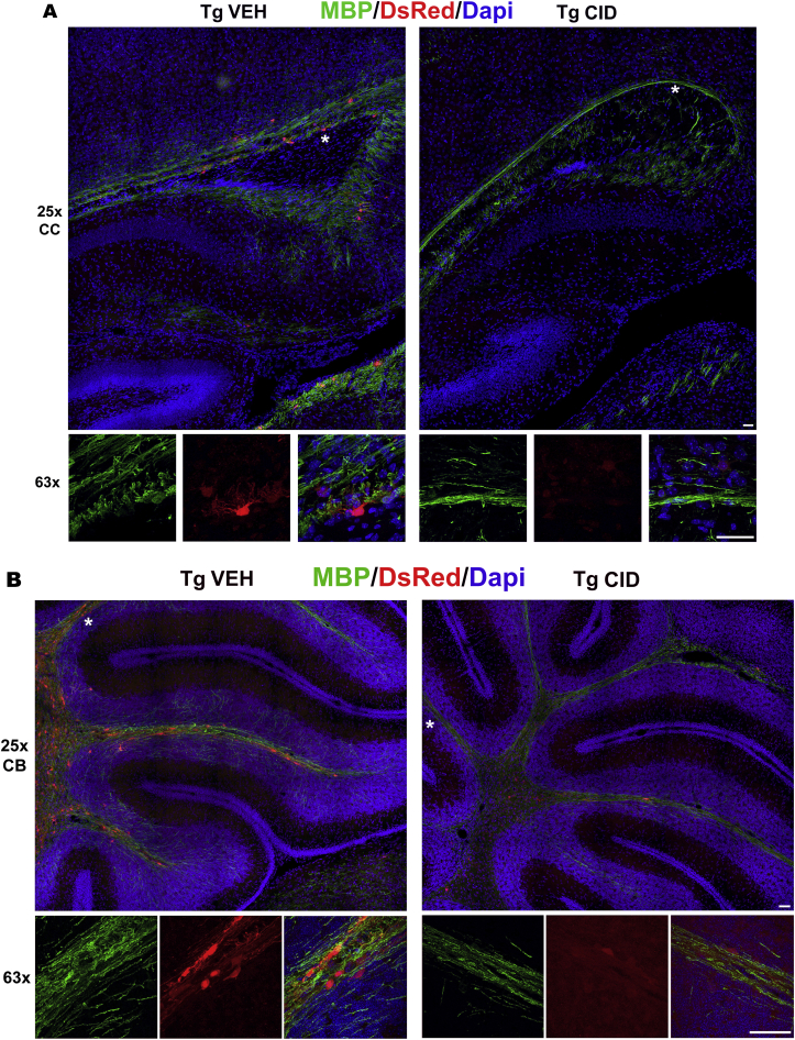
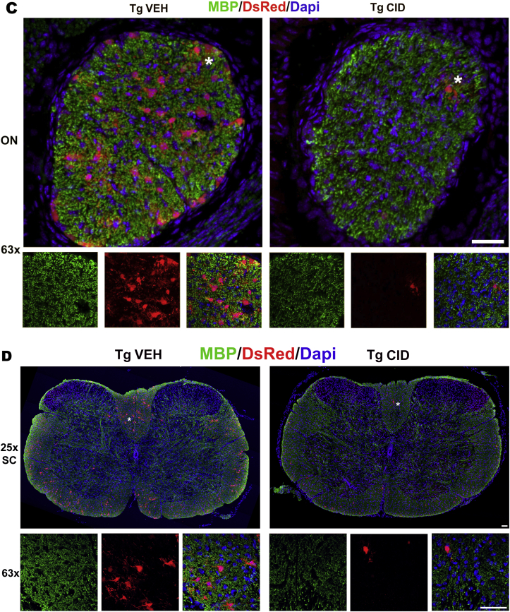
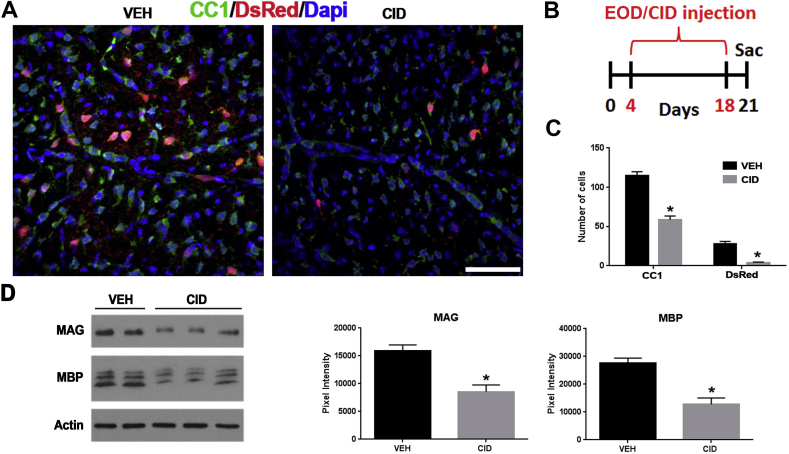
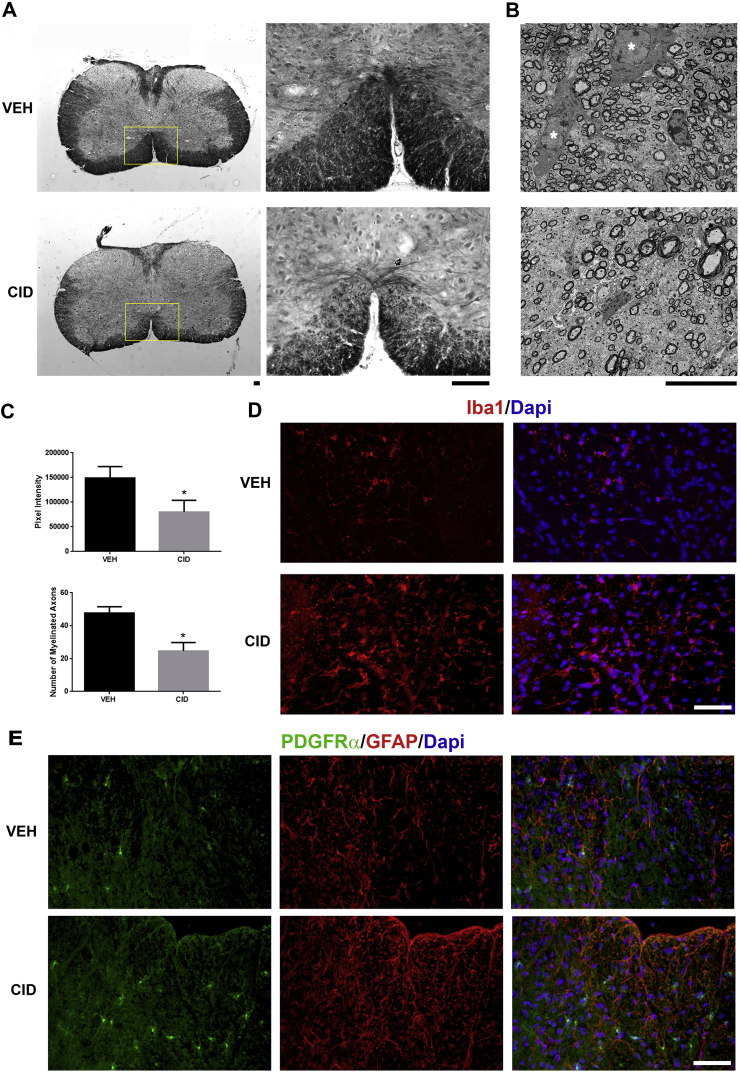
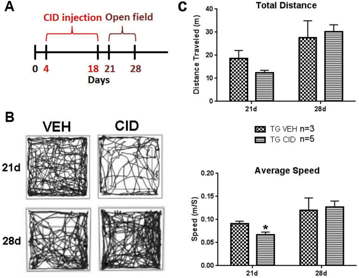
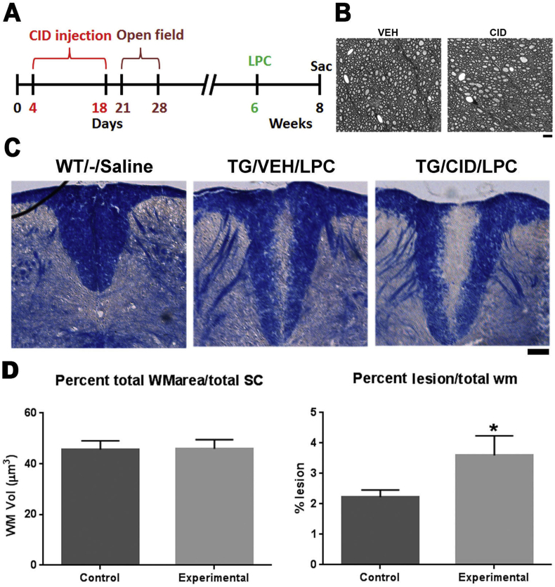
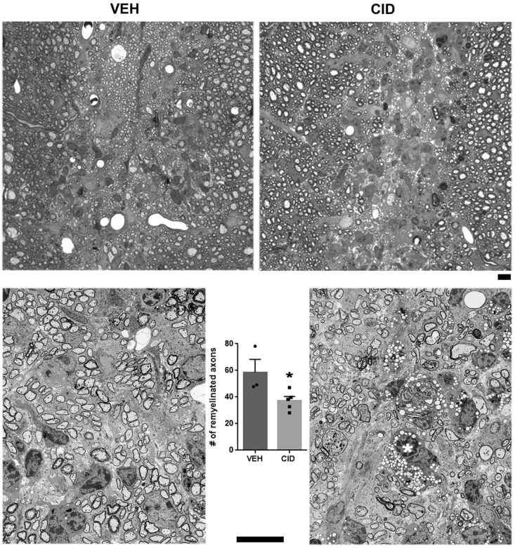
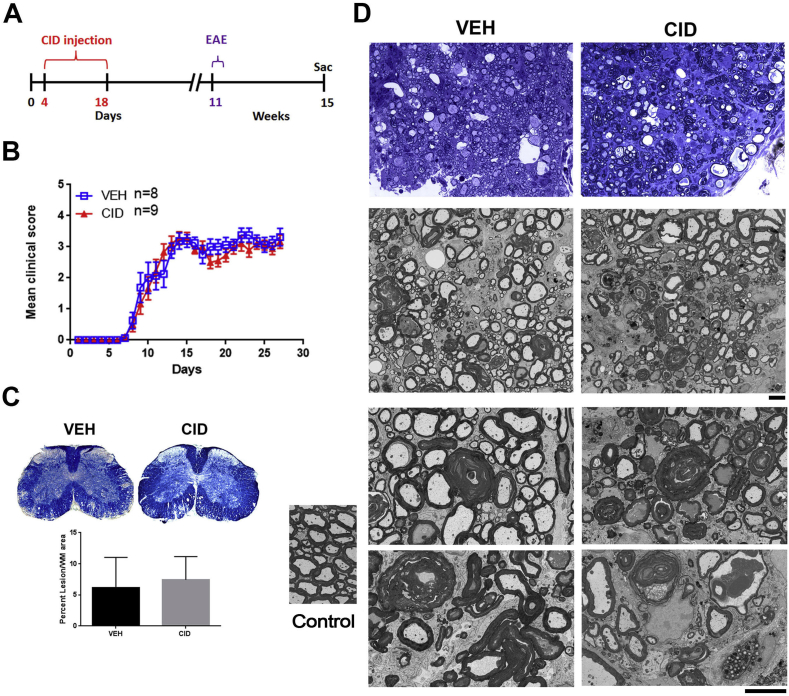
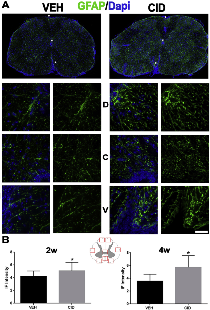
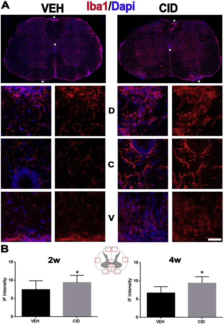
Multiple sclerosis (MS) is a CNS neurodegenerative autoimmune disease characterized by loss of oligodendrocytes and myelin in the brain and the spinal cord that results in localized functional deficits. Several risk factors have been associated with MS, however none fully explain the enhanced susceptibility seen in older individuals. Epidemiological data, based on geographical prevalence studies suggest that susceptibility is established early in life and frequently long before the diagnosis of disease raising the possibility that developmental events influence adult disease onset and progression. Here we test the hypothesis that selective loss of mature oligodendrocytes during postnatal development results in enhanced susceptibility to a demyelinating insult to the mature CNS. A transgenic mouse model was utilized to specifically induce apoptotic cell death in a subset of mature oligodendrocytes (MBP-iCP9) during the first 2 postnatal weeks followed by either a local LPC spinal cord injection or the induction of EAE in the adult animal. Immunostaining, immunoblotting, behavioral testing, and electron microscopy were utilized to examine the differences in the response between animals with developmental loss of oligodendrocytes and controls. We show that during development, oligodendrocyte apoptosis results in transient reductions in myelination and functional deficits that recover after 10-14 days. Compared to animals in which oligodendrocyte development was unperturbed, animals subjected to postnatal oligodendrocyte loss showed delayed recovery from an LPC lesion to the mature spinal cord. Unexpectedly, the induction and severity of MOG induced EAE was not significantly altered in animals following oligodendrocyte developmental loss even though there was a substantial increase in spinal cord tissue damage and CNS inflammation. It is unclear why the elevated glial responses seen in developmentally compromised animals were not reflected in enhanced functional deficits. These observations suggest that developmental loss of oligodendrocytes results in long lasting tissue changes that alter its response to subsequent insults and the capacity for repair in the adult.

Keywords: EAE; Inflammation; LPC; Multiple sclerosis; Priming.


Conflict of interest statement

We do not have any conflict of interest regarding this study.

Figures

Fig. 1
Fig. 1
Systemic CID injection induces loss of DsRed ​+ oligodendrocytes and a decrease in MBP staining throughout the CNS. Sections of tissues taken 3 days after the final injection of CID (P4–P11) show reduction in the number of oligodendrocytes and myelin throughout the CNS. Corpus callosum (A), cerebellum (B), optic nerve (C), and spinal cord (D) representative sections from MBP-iCP9 transgenic mice subcutaneously injected with vehicle (VEH) or CID (n ​= ​3/group) were labeled with antibodies to MBP (green), DsRed (red), and DAPI (blue). The bottom panel in each image shows the areas indicated with an asterisk in the top panel (Bar ​= ​25 ​μm). (For interpretation of the references to colour in this figure legend, the reader is referred to the Web version of this article.)
Fig. 1
Fig. 1
Systemic CID injection induces loss of DsRed ​+ oligodendrocytes and a decrease in MBP staining throughout the CNS. Sections of tissues taken 3 days after the final injection of CID (P4–P11) show reduction in the number of oligodendrocytes and myelin throughout the CNS. Corpus callosum (A), cerebellum (B), optic nerve (C), and spinal cord (D) representative sections from MBP-iCP9 transgenic mice subcutaneously injected with vehicle (VEH) or CID (n ​= ​3/group) were labeled with antibodies to MBP (green), DsRed (red), and DAPI (blue). The bottom panel in each image shows the areas indicated with an asterisk in the top panel (Bar ​= ​25 ​μm). (For interpretation of the references to colour in this figure legend, the reader is referred to the Web version of this article.)
Fig. 2
Fig. 2
Oligodendrocyte ablation results in a significant decrease in mature oligodendrocytes and a decrease in myelin proteins. A. Representative images of spinal cord sections labeled with antibodies to CC1 (green), DsRed (red), and DAPI (blue) in vehicle (VEH) and CID injected pups. Note that the CID injected animals (n ​= ​2) show a significant decrease in CC1 and DsRed staining as compared to vehicle. B. Schematic of the every other day (EOD) CID injection and tissue collection. C. Quantification of cell numbers indicate a significant decrease in the number of mature CC1+ oligodendrocytes (p-value ​= ​0.0312) and DsRed ​+ ​cells (p-value ​= ​0.0001) in CID injected, compared to VEH injected animals. CC1+ and DsRed ​+ ​cells were counted from 6 areas (2 dorsal, 2 ventral, and 2 lateral) of the spinal cord. D. Western blot analysis of spinal cord tissue indicates that the myelin proteins MAG (p-value ​= ​0.007) and MBP (p-value ​= ​0.005) were decreased significantly after CID injection compared to VEH treated animals (CID n ​= ​3 and VEH n ​= ​2) (Bar ​= ​25 ​μm). (For interpretation of the references to colour in this figure legend, the reader is referred to the Web version of this article.)
Fig. 3
Fig. 3
Developmental ablation of mature oligodendrocytes results in a decrease in myelinated axons, an increase in astrocytes and microglial activation, and elevated numbers of oligodendrocyte progenitor cells. A. Representative images of spinal cord sections stained with Solochrome indicate a decrease in myelin staining observed throughout the white matter in animals treated with CID (n ​= ​3) compared to vehicle (VEH) treated controls (n ​= ​2). Yellow boxes represent magnified areas. B. Ultrastructural analyses confirmed the decrease in myelinated axons in CID compared to VEH treated spinal cord. Healthy oligodendrocytes (white asterisk) are apparent in the VEH image associated with neighboring axons. C. Quantification of Solochrome images (top graph) and EM sections (bottom graph) show significant decrease in levels of myelin and the number of myelinated axons in CID compared to VEH animals. D, E. Oligodendrocyte ablation results in changes in adjacent glial cell populations. Sections from spinal cord injected with VEH or CID were labeled with antibodies to Iba1 (red in D), PDGFRα (green in E), GFAP (red in E), and Dapi (blue). CID treated animals demonstrated increased Iba1 (D) and GFAP (E) immunoreactive and higher number of OPCs compared to VEH animals. (Bar ​= ​10 ​μm in A and B, Bar ​= ​25 ​μm in D and E). (For interpretation of the references to colour in this figure legend, the reader is referred to the Web version of this article.)
Fig. 4
Fig. 4
Oligodendrocytes ablation results in a transient functional deficit. A. Schematic of the CID injection and open filed testing. B. Representative traces obtained from mice at P21 (3 days post-CID) and P28 (10 days post-CID) (vehicle/VEH n ​= ​3, CID n ​= ​5). At P21, the motility of the animals is significantly reduced in CID treated animals compared to VEH treated controls as indicated by the density of traces. At P28 the level of motility between the two groups was similar. C. Quantification of the differences in total distance and average speed traveled indicates a decrease in total distance and a significant decrease (p-value ​= ​0.0316) in average speed traveled by CID treated mice at 3 days post-CID (21 days) that was resolved by10 days post-CID (28 days).
Fig. 5
Fig. 5
Larger LPC-induced demyelinated lesionsare presentin CID treated mice than controls. A. Schematic of the CID injection and the time-line for LPC injections. B. Representative EM images from the ventral regions show no significant differences in the number of myelinated axons and myelin thickness between VEH and CID treated animals at 8 weeks of age. C. Representative images show sections from wild type (WT) mouse injected with saline (WT/-/Saline), MBP-iCP9 transgenic mouse injected with vehicle (VEH) followed by LPC (TG/VEH/LPC), and MBP-iCP9 transgenic mouse injected with CID followed by LPC (TG/CID/LPC). Sections were taken 14 days after LPC injection. WT mice injected with saline did not show any detectable lesion. WT/CID/LPC and TG/VEH/LPC treated mice (Controls) served as control groups in which oligodendrocytes were not ablated. D. The total volume of white matter (WM) area was compared to the total volume of spinal cord across all groups and no significant differences were detected suggesting that normal myelination was unaffected by creation of the transgenic phenotype. Two sections from each animal were assessed and the relative proportion of lesioned dorsal white matter was significantly (p-value ​= ​0.0039) higher in experimental transgenic mice, that had received CID and LPC (n ​= ​3) compared to those that received vehicle and LPC or wild type mice that received CID (n ​= ​2) suggesting slower remyelination in the CID treated transgenic mice (Bar in B ​= ​25 ​μm).
Fig. 6
Fig. 6
Remyelination is reduced in LPC-induced demyelination in CID treated transgenic mice. Vehicle (VEH, n ​= ​3) and CID treated (n ​= ​5) MBP-iCP9 transgenic mice were injected with LPC at 6 weeks (about 4 weeks after the last CID injection) and sacrificed at 8 weeks of age. Sections were stained using Toluidine Blue stain (upper panels) and remyelinated axons were counted in three regions in the dorsal column. There is a significant decrease (p-value ​= ​0.0225) in the number of remyelinated axons indicating decreased recovery in the CID treated mice. Lower panels show the SEM images. Note the lower level of myelinated axons and the presence of foamy macrophages in the CID treated tissue as indicated by the asterisk (Bar ​= ​10 ​μm).
Fig. 7
Fig. 7
CID treated animals have worse histological outcomes in EAE,which is not reflected in the functional outcome. A. Schematic of the CID injection and the timeline for EAE induction. B. Both Vehicle (VEH, n ​= ​8) and CID (n ​= ​9) treated mice were induced with EAE at 11 weeks of age (about 8 weeks after CID injection). There are no significant differences in the severity of the disease as indicated by the mean clinical score (B) or the overall level of lesion load based on Solochrome staining (C). However, the tissue integrity (D) was worse in the CID treated with a greater degree of myelin perturbation and axonal degeneration (Bar ​= ​5 ​μm).
Fig. 8
Fig. 8
Astrocyte activation is increased in the spinal cord of animals with EAE that were subjected to developmental ablation of mature oligodendrocytes. A. Sections from spinal cord of EAE mice treated developmentally with vehicle (VEH, n ​= ​2) or CID (n ​= ​2) were labeled with antibodies to GFAP (green) and Dapi (blue) 2 weeks after EAE induction. Bottom panels show higher magnifications (63×) of the areas indicated by asterisks in the lower magnification (25×) images in the top panels. The higher magnification areas are taken from (top to bottom) the dorsal (D), near the central canal (C), and ventral (V) spinal cord. B. Intensity of immunofluorescent staining was measured using image J in 8 different areas from each mouse, as depicted in the cartoon. Astrocytes are significantly more activated in the CID treated tissues at 2 (p-value ​= ​0.0364) and 4 weeks (p-value ​= ​0.0003) after EAE induction (Bar ​= ​25 ​μm in A). (For interpretation of the references to colour in this figure legend, the reader is referred to the Web version of this article.)
Fig. 9
Fig. 9
Microglial activation is increased in the spinal cord of animals with EAE subjected to developmental ablation of mature oligodendrocytes. A. Sections from spinal cord of EAE mice treated with vehicle (VEH, n ​= ​2) or CID (n ​= ​2) were labeled with antibodies to Iba1 (red) and Dapi (blue) 2 weeks after EAE induction. Bottom panels show higher magnifications (63×) of the areas indicated by asterisks in the lower magnification (25×) images. The higher magnification areas are taken from (top to bottom) the dorsal (D), near the central canal (C), and ventral (V) spinal cord. The increase in the number and morphology of the microglia is evident in the CID treated tissues as indicated by the change in the staining intensity and the morphology of the microglia from more ramified in the VEH treated to more amoeboid in the CID treated tissues. B. Intensity of immunofluorescent staining was measured using image J in 8 different areas from each mouse, as depicted in the cartoon. Microglia are significantly more activated in the CID treated tissues at 2 (p-value ​= ​0.0161) and 4 weeks (p-value ​= ​0.0001) after EAE induction as indicated by increase in Iba1 labeling intensity and increased adoption of an amoeboid morphology (Bar ​= ​25 ​μm in A). (For interpretation of the references to colour in this figure legend, the reader is referred to the Web version of this article.)

Similar articles

Cited by

References

    1. Bilbo S.D. Neonatal infection induces memory impairments following an immune challenge in adulthood. Behav. Neurosci. 2005;119(1):293–301. - PubMed
    1. Boisse L. Long-term alterations in neuroimmune responses after neonatal exposure to lipopolysaccharide. J. Neurosci. 2004;24(21):4928–4934. - PMC - PubMed
    1. Caprariello A.V. Apoptosis of oligodendrocytes in the central nervous system results in rapid focal demyelination. Ann. Neurol. 2012;72(3):395–405. - PMC - PubMed
    1. Caprariello A.V. Apoptosis of oligodendrocytes during early development delays myelination and impairs subsequent responses to demyelination. J. Neurosci. 2015;35(41):14031–14041. - PMC - PubMed
    1. Cunningham C. Systemic inflammation induces acute behavioral and cognitive changes and accelerates neurodegenerative disease. Biol. Psychiatr. 2009;65(4):304–312. - PMC - PubMed

LinkOut - more resources