Observational Study
. 2022 Aug;35(4):527-532.
doi: 10.1177/19714009211049089. Epub 2021 Sep 30.

Cone-beam CT angiography to assess the microvascular anatomy of intracranial arterial dissections

Affiliations
Observational Study

Cone-beam CT angiography to assess the microvascular anatomy of intracranial arterial dissections

Francesco Diana et al. Neuroradiol J. 2022 Aug.

Abstract

Background: Intracranial artery dissection is a rare and generally under-recognized cause of ischaemic stroke or subarachnoid haemorrhage.

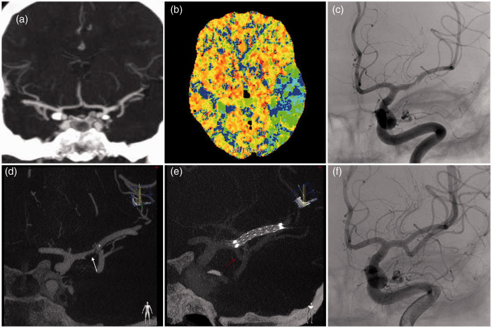
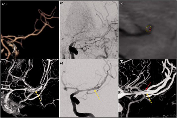
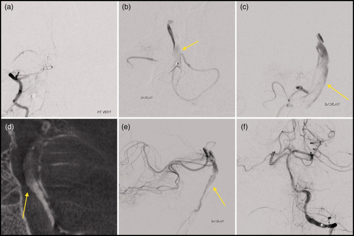
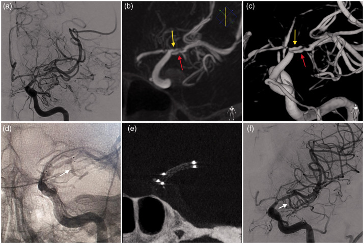
Objectives: The aim of this study was to analyse the efficacy of cone-beam computed tomography angiography (CBCT-A) to detect arterial ultrastructural alterations in intracranial artery dissection.

Method: This is an observational and retrospective case series.

Results: Between January 2018 and November 2020, four patients were admitted with an acute ischaemic stroke due to intracranial dissection studied with CBCT-A. In all cases, the CBCT-A documented vascular ultrastructural alterations related with the intracranial dissection.

Conclusions: CBCT-A is an intraprocedural diagnostic technique that is useful for the diagnosis of intracranial dissections.

Keywords: Cone beam CT; intracranial dissection; intracranial stent; stroke; thrombectomy.


Figures

Figure 1.
Figure 1.
Patient 1. (a) Computed tomography angiography showing a filling defect with stenosis in the distal left M1 segment and altered flow in the inferior division. (b) Magnetic resonance perfusion showing increased mean transit time (MTT) in the left temporal lobe. (c) Left internal carotid artery frontal angiographic run demonstrating stenosis of the left middle cerebral artery (MCA) and occlusion of the inferior division. (d) First cone-beam computed tomography angiography (CBCT-A) showing the mural haematoma or a clot into the false lumen (white arrow) and the intimal flap (white asterisk). (e) and (f) Second CBCT-A and control angiogram depicting apposition of the intracranial stent and recanalized inferior division (red arrow).
Figure 2.
Figure 2.
Patient 2. (a) Computed tomography angiography done a few months before the event, which showed normal left middle cerebral artery (MCA). (b) Left internal carotid artery frontal angiographic run showing the narrowing of the MCA. (c) Magnetic resonance imaging with sagittal T1 showing the mural haematoma (yellow circle) and the true lumen (red circle). (d) Cone-beam computed tomography angiography (CBCT-A) performed one week later documenting the dissection with partially recanalized false lumen (yellow arrow). (e) and (f) One year follow-up angiogram and CBCT-A showing the filiform true lumen (red arrow) giving rise only to a lateral lenticulostriate arteries and the recanalized false lumen (yellow arrow) which supplies both divisions.
Figure 3.
Figure 3.
Patient 3. (a) Angiographic run from right vertebral artery confirming occlusion of the proximal basilar artery. (b) and (c) Frontal and lateral projections from microcatheter injection of the proximal basilar artery after aspiration showing spiraliform filling of a false lumen of the basilar artery (yellow arrow). (d) to (f) CBCT-A and angiographic run showing antegrade filling of the basilar artery false lumen (yellow arrow) emptying into the true lumen distally without antegrade filling of the cerebellar arteries.
Figure 4.
Figure 4.
Patient 4. (a) Angiographic run showing an irregular appearance of the proximal left middle cerebral artery (MCA). (b/c) Cone-beam computed tomography angiography (CBCT-A) depicted in detail the dissection, with a transverse flap across the MCA dividing a false lumen (yellow arrow) and the true lumen (red arrow). (d) to (f) Angiographic control and CBCT-A at the end of the treatment showing the apposition of the intracranial stent and the recanalized temporal branch (white arrow).

Similar articles

References

    1. Anson J, Crowell RM. Cervicocranial arterial dissection. Neurosurgery 1991; 29: 89–96. - PubMed
    1. Thanvi B, Munshi SK, Dawson SL, et al.. Carotid and vertebral artery dissection syndromes. Postgrad Med J 2005; 81: 383–388. - PMC - PubMed
    1. Ohkuma H, Suzuki S, Ogane K; Study Group of the Association of Cerebrovascular Disease in Tohoku, Japan. Dissecting aneurysms of intracranial carotid circulation. Stroke 2002; 33: 941–947. - PubMed
    1. Moonis G, Hwang CJ, Ahmed T, et al.. Otologic manifestations of petrous carotid aneurysms. AJNR Am J Neuroradiol 2005; 26: 1324–1327. - PMC - PubMed
    1. Labeyrie M-A, Civelli V, Reiner P, et al.. Prevalence and treatment of spontaneous intracranial artery dissections in patients with acute stroke due to intracranial large vessel occlusion . J Neurointerv Surg 2018; 10: 761–4. - PubMed

Publication types