. 2022 Jan 13;106(1):155-172.
doi: 10.1093/biolre/ioab183.

Single-cell analysis of prostaglandin E2-induced human decidual cell in vitro differentiation: a minimal ancestral deciduogenic signal†

Single-cell analysis of prostaglandin E2-induced human decidual cell in vitro differentiation: a minimal ancestral deciduogenic signal†

Daniel J Stadtmauer et al. Biol Reprod. .

Abstract

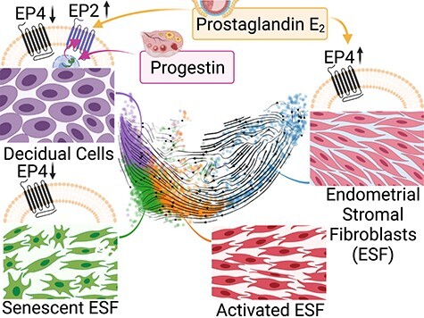
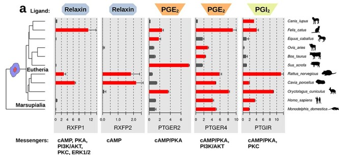
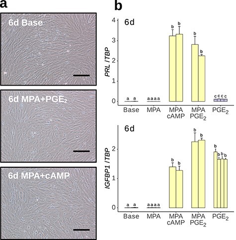
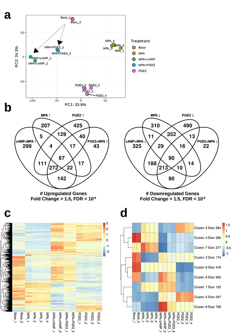
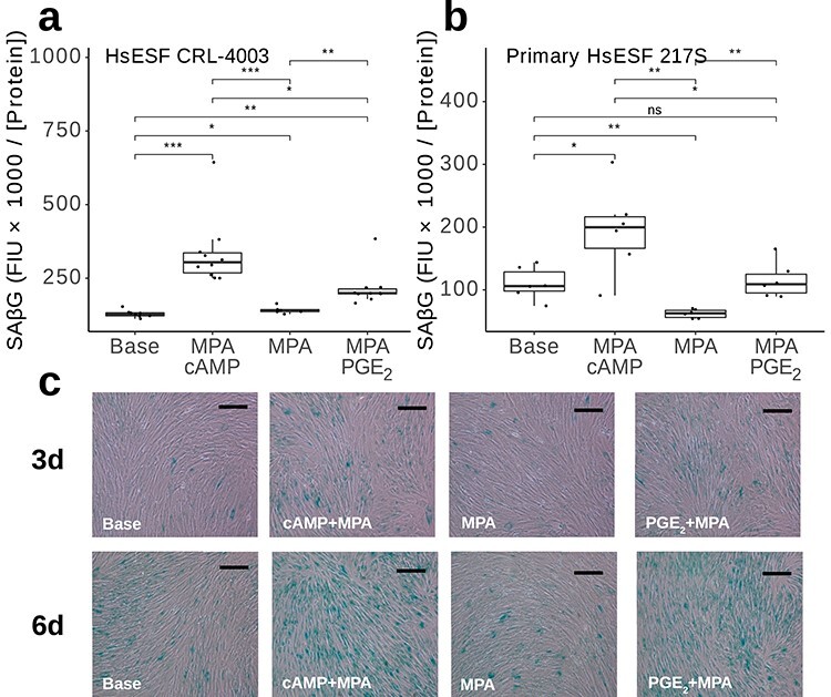
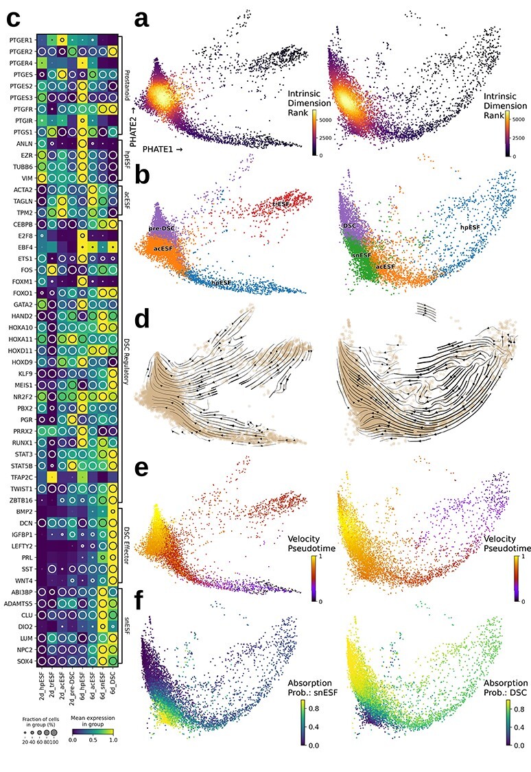
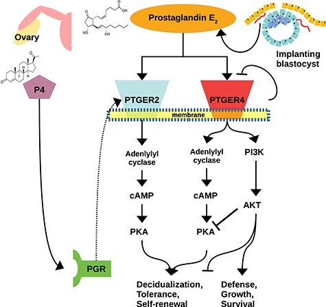
The decidua is a hallmark of reproduction in many placental mammals. Differentiation of decidual stromal cells is known to be induced by progesterone and the cyclic AMP/protein kinase A (cAMP/PKA) pathway. Several candidates have been identified as the physiological stimulus for adenylyl cyclase activation, but their relative importance remains unclear. To bypass this uncertainty, the standard approach for in vitro experiments uses membrane-permeable cAMP and progestin. We phylogenetically infer that prostaglandin E2 (PGE2) likely was the signal that ancestrally induced decidualization in conjunction with progesterone. This suggests that PGE2 and progestin should be able to activate the core gene regulatory network of decidual cells. To test this prediction, we performed a genome-wide study of gene expression in human endometrial fibroblasts decidualized with PGE2 and progestin. Comparison to a cAMP-based protocol revealed shared activation of core decidual genes and decreased induction of senescence-associated genes. Single-cell transcriptomics of PGE2-mediated decidualization revealed a distinct, early-activated state transitioning to a differentiated decidual state. PGE2-mediated decidualization was found to depend upon progestin-dependent induction of PGE2 receptor 2 (PTGER2) which in turn leads to PKA activation upon PGE2 stimulation. Progesterone-dependent induction of PTGER2 is absent in opossum, an outgroup taxon of placental mammals which is incapable of decidualization. Together, these findings suggest that the origin of decidualization involved the evolution of progesterone-dependent activation of the PGE2/PTGER2/PKA axis, facilitating entry into a PKA-dominant rather than AKT-dominant cellular state. We propose the use of PGE2 for in vitro decidualization as an alternative to 8-Br-cAMP.

Keywords: PTGER2; cell type origination; decidua; decidual stromal cell; endometrial stromal fibroblast; evolution of mammalian pregnancy; prostaglandin E2; senescence.


Figures

Graphical Abstract
Graphical Abstract
Figure 1
Figure 1
Expression of receptors for potential deciduogenic ligands in unstimulated primary ESF from 11 species. RLN receptors: RXFP1, RXFP2; PGE2 receptors: PTGER2, PTGER4; and prostaglandin I2 receptor: PTGIR. Data from Ma et al. [7] depicted as mean square-root TPM from all replicates. Error bars represent standard error of the mean. Tree topology from dos Reis et al. [90].
Figure 2
Figure 2
In vitro decidualization of human ESF using PGE2 + MPA. (a) Cells treated with culture media for 6 days (6d Base) retained spindle morphology, whereas cells treated with MPA plus PGE2 or cAMP for 6 days became more globular. Scale bar = 20 μm. (b) qPCR quantification of decidual markers PRL and IGFBP1 in response to various decidualization regimes, measured as linearized expression relative to control gene TBP. Technical n = 3 for all conditions.
Figure 3
Figure 3
Bulk RNA sequencing analysis. (a) Principal component analysis conducted on protein-coding genes. Transcriptomes cluster by treatment. Arrows drawn represent putative decidualization trajectories. (b) Venn diagrams of genes differentially upregulated or downregulated in response to cAMP + MPA or PGE2 + MPA treatments with respect to control treatments. (c) Heatmap of differentially expressed protein-coding genes. (d) K-means clustered (k = 9) heatmap of differentially expressed protein-coding genes showing major patterns of comparative gene expression among the treatment conditions.
Figure 4
Figure 4
SAβG assays. (a, b) Fluorimetric quantification of SAβG in Fluorescence Intensity Units (FIU) divided by protein concentration in μg/μL for major treatments plus 250 nM dasatinib (Das) have been indicated by using immortalized (a) and primary (b) human ESF. t-test P values: ns: P > 0.05; *: P ≤ 0.05; **  P ≤ 0.01; ***: P ≤ 0.001. (c) Representative images of chromogenic SAβG assays on immortalized human ESF treated for 3 and 6 days as indicated. Scale bars = 20 μm.
Figure 5
Figure 5
Single-cell clusters and their differentially expressed genes. (a) PHATE embedding of 2-day (left) and 6-day (right) DSC colored by intrinsic dimension (ordinal). (b) Labeled k-means cell clusters. (c) Normalized expression values of marker genes for identified clusters, with enclosed circle size corresponding to the percentage of cells expressing the gene at greater than 1 CPM. (d) RNA velocity stream embedding showing velocity-inferred direction of cellular state transition. (e) Velocity-informed pseudotime of cells showing progression toward. (f) Absorption probabilities for senescent (left) and mature (right) decidual end-points identified at day 6.
Figure 6
Figure 6
PTGER2 expression is upregulated by progestin and contributes to PGE2-induced decidualization. (a) Expression of PTGER2 (top) and PTGER4 (bottom) relative to latent time pseudotemporal progression toward DSC or snESF in 6-day single-cell data and colored by absorption probability to either DSC or snESF macrostates. Plots to the right show expression on PHATE ordinations. (b) qPCR of PRL in response to PGE2 + MPA decidualization plus inhibitors of PTGER2 (EP2i) or PTGER4 (EP4i), or PKA (PKAi). (c) Gene expression of PGE2 receptors 2 and 4 in human neo-ESF (top) and Monodelphis domestica paleo-ESF (bottom; data from [6]). (d–f) qPCR quantification of PTGER2 after 8–24 h (d), 48 h (e), or 6 days (f) indicated treatment. (g) Overall expression scores for AKT-related genes in 6-day DSC.
Figure 7
Figure 7
Schematic diagram of induction of decidual cell differentiation. Self-limitation of PTGER4, plus P4-induced transcriptional upregulation of PTGER2, result in a transition to a PKA-dominated, rather than AKT-dominated, signaling state.

References

    1. Gellersen B, Brosens JJ. Cyclic decidualization of the human endometrium in reproductive health and failure. Endocr Rev 2014; 35:851–905. - PubMed
    1. Mess A, Carter AM. Evolutionary transformations of fetal membrane characters in Eutheria with special reference to Afrotheria. J Exp Zool Part B 2006; 306:140–163. - PubMed
    1. Chavan AR, Griffith OW, Wagner GP. The inflammation paradox in the evolution of mammalian pregnancy: turning a foe into a friend. Curr Opin Genet Dev 2017; 47:24–32. - PubMed
    1. Chavan AR, Griffith OW, Stadtmauer DJ, Maziarz J, Pavličev M, Fishman R, Koren L, Romero R, Wagner GP. Evolution of embryo implantation was enabled by the origin of decidual cells in eutherian mammals. Mol Biol Evol 2021; 38:1060–1074. - PMC - PubMed
    1. Stadtmauer DJ, Wagner GP. The primacy of maternal innovations to the evolution of embryo implantation. Integr Comp Biol 2020; 60:742–752. - PMC - PubMed

Publication types

MeSH terms