. 2021 Oct;374(6563):eabf2911.
doi: 10.1126/science.abf2911. Epub 2021 Oct 1.

A protein network map of head and neck cancer reveals PIK3CA mutant drug sensitivity

Affiliations

A protein network map of head and neck cancer reveals PIK3CA mutant drug sensitivity

Danielle L Swaney et al. Science. 2021 Oct.

Abstract

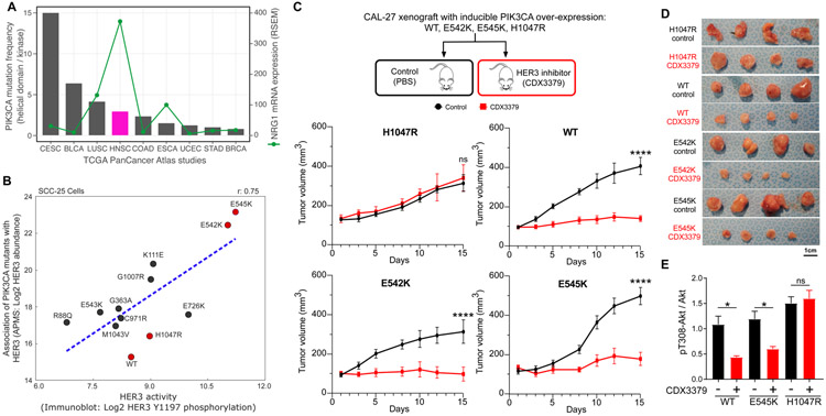
We outline a framework for elucidating tumor genetic complexity through multidimensional protein-protein interaction maps and apply it to enhancing our understanding of head and neck squamous cell carcinoma. This network uncovers 771 interactions from cancer and noncancerous cell states, including WT and mutant protein isoforms. Prioritization of cancer-enriched interactions reveals a previously unidentified association of the fibroblast growth factor receptor tyrosine kinase 3 with Daple, a guanine-nucleotide exchange factor, resulting in activation of Gαi- and p21-activated protein kinase 1/2 to promote cancer cell migration. Additionally, we observe mutation-enriched interactions between the human epidermal growth factor receptor 3 (HER3) receptor tyrosine kinase and PIK3CA (the alpha catalytic subunit of phosphatidylinositol 3-kinase) that can inform the response to HER3 inhibition in vivo. We anticipate that the application of this framework will be valuable for translating genetic alterations into a molecular and clinical understanding of the underlying biology of many disease areas.


Figures

Figure 1.
Figure 1.. Experimental design and workflow.
(A) The alteration frequencies from the HNSCC TCGA provisional dataset (n = 530 patients) for the 31 experimentally tractable genes selected as AP-MS baits in this study. Proteins analyzed in this study are listed, along with the percentage of patients with an alteration in that gene/protein. Each patient is represented by a grey box that is colored based on the occurrence and type of alteration(s) observed in that patient. Both the wild-type and mutant protein sequence(s) were analyzed for genes highlighted in yellow. The genetic alteration types in the two cancer cell lines (CAL-33 and SCC-25) are also displayed. (B) The experimental workflow in which each bait was expressed in biological triplicate in 3 cell lines and subjected to AP-MS analysis. (C-E) Permutation test illustrating the frequency of CNVs (C), mRNA alterations (D), or mutations (E), from randomly selected genes in the HNSCC TCGA data. The white circle indicates the median of the random sampling, and the grey bar represents +/− 1 standard deviation. The frequency of alterations found in the prey retrieved in this PPI dataset is indicated in the black circle. (F) Percentage of HC-PPIs identified in a panel of public PPI databases (CORUM, BioPlex 2.0, or BioGRID low throughput and multivalidated, and IMEX (23, 36-38)). (G) Clustering analysis of all HC-PPIs (n = 771) based on their PPI score, which is an average of the confidence scores reported from SAINTexpress and CompPASS score (see Materials and methods for details). A PPI score of 1.0 represents the highest confidence in a PPI. (H) Venn diagram illustrating the overlap in HC-PPIs among the 3 cell lines. For this analysis, only those PPIs passing the HC-PPI filtering criteria by both SAINTexpress and CompPASS were classified as an HC-PPI within an individual cell line.
Figure 2.
Figure 2.. Differential interaction analysis of the HNSCC enriched and depleted interactome.
(A) Interactome of the union of all HC-PPIs detected across all cell lines. Edges are colored based on their differential interaction score (DIS), with pink edges representing PPIs that are enriched in HNSCC (both SCC-25 and CAL-33) as compared to HET-1A cells, and teal lines representing PPIs that are depleted from HNSCC cell lines. IAS connections represent physical protein-protein association derived from prior studies (55) (see Material and methods). (B) For baits with ∣DIS∣ > 0.5, the fraction of PPIs for that bait having HNSCC-enriched PPIs with DIS > 0.5, or HNSCC-depleted DIS < −0.5. (C) CCND1 interactome. Here the SAINTexpress score, used for calculation of the DIS, is displayed for each cell line within the prey node, ND indicates not detected. (D) DIS for the entire interactome represented in panel A ranked by DIS. (E) Subnetwork of the interactome of the HNSCC-enriched and -depleted interactions.
Figure 3.
Figure 3.. An HNSCC-enriched FGFR3:Daple interaction mediates activation of cell migratory proteins.
(A) Differential scoring analysis of the FGFR3 interactome highlights CCDC88C (Daple) as an HNSCC-enriched interaction partner to both FGFR3 and ERBB2 (HER2). (B) Activation of RTKs can disrupt the interaction between Disheveled (Dvl) and Daple, allowing Daple to function as a GEF for Gαi. GTP binding causes dissociation of the G protein, leaving Gβγ subunits free to activate migratory signaling through Rac and PAK. (C) NanoBiT biosensor measures Gαi activation through dissociation of the luciferase split between Gα and Gβγ. CNO mediates canonical GPCR signaling through the synthetic Gαi-coupled DREADD receptor. FGF mediates HNSCC-specific signaling through FGFR3 and Daple. (D) Luminescence was measured in CAL-33 and HET-1A cells transfected with Gαi NanoBiT and siRNA (control, FGFR3, or Daple) and stimulated with FGF (10ng/mL) (*P < 0.05 when compared with the vehicle-treated group). (E) Immunoblot analysis of CAL-33 subject to siRNA knockdown. (F) PAK1/2 autophosphorylation measured by immunoblot analysis over a time course of FGF stimulation (0, 5, 10, 30, 60 minutes) in CAL-33 and HET-1A cells. (G) PAK1/2 autophosphorylation measured by immunoblot analysis in CAL-33 cells stimulated with FGF (10ng/mL) and/or treated with 0.5μM of the pan FGFR inhibitor Infigratinib (*P < 0.05 when compared with the vehicle-treated group). (H-I) A vertical scratch was introduced to fibronectin-plated CAL-33 cells and cells were stimulated with FGF (10ng/mL) and/or treated with 0.5μM of Infigratinib. Replicate scratch closures were quantified (H, (*P < 0.05, **P < 0.01 when compared with the vehicle-treated group)), and (I) images were taken at 0 and 24 hours after FGF stimulation (scale bar = 250μm). (J) Daple and FGFR3 expression are plotted for all upper airway and esophageal cell lines in DepMap (62), with the two cancer cell lines used in this study highlighted in red. (K) The sensitivity of cell lines with high or low Daple expression to either a FGFR1 inhibitor (sorafinib), or a FGFR1/2/3 inhibitor (AZD4547) as quantified by area under the curve (AUC) (*P < 0.05). Cell lines were selected from panel J, and for those with corresponding drug sensitivity data the top 5 Daple expressing cells (High Daple) or the bottom 5 Daple expressing cells (Low Daple), were used.
Figure 4.
Figure 4.. PIK3CA mutant interactome.
(A) Overview of the PIK3CA signaling pathway, which is often stimulated by RTKs that interact with PIK3CA to stimulate RAS/Raf-mediated or Akt/mTORC1-mediated downstream signaling. (B) Analyzed PIK3CA mutants and their frequency in HNSCC tumors from TCGA. Asterisk (*) denotes mutations annotated as oncogenic in OncoKB (65). Graph bars corresponding to each mutation were color-coded to indicate their localization within the p110α domain (as indicated in the legend in top right corner). (C) Selected PIK3CA mutations were mapped on the structure of PI3K (PDB: 4L23) (66) by highlighting the mutated residues as red spheres. (D) Quantification of PPIs for all PIK3CA HC-PPIs detected in the SCC-25 cell line (all cell lines displayed in Figure S4A). (E) Cartoon representation of a zoomed-in view of PI3K illustrating a salt bridge formed between K11 and E81 (PDB: 4L23). (F) A zoomed-in view depicting interactions made by G1007 in PI3K (PDB: 4L23). (G) Cartoon representation of different mutation induced PI3K activation mechanisms and their respective HER3 binding preferences.
Figure 5.
Figure 5.. In vivo targeting of HER3 in the context of different PIK3CA mutants.
(A) Bar chart representing the ratio of helical domain (E545 and E542) mutations as compared to kinase domain mutations (H1047) across TCGA PanCancer Altas studies represented in cBioPortal (68). Line graph showing the mRNA expression (RSEM) for NRG1 across the same studies. (B) Correlation of Log2 HER3 interaction levels from AP-MS experiments and Log2 HER3 Y1197 phosphorylation levels from immunoblot analysis. All values are normalized by FLAG-PIK3CA levels in their respective experiments. Mutations marked in red were selected for in vivo experiments. (C-D) CAL-27 cells expressing inducible PIK3CA variants were transplanted into athymic nude mice. Mice were fed with doxycycline to induce PIK3CA expression. When tumor volumes reached approximately 100 mm3, mice were treated with vehicle (PBS) or CDX3379 (10mg/kg, twice a week) for approximately 15 days, as indicated. Shown are (C) tumor growth curves, (D) representative tumor images, and (C) last day tumor volume (****P < 0.0001 when compared with the control-treated group). (E) Quantification of immunoblot analysis of signaling events in the same CAL-27 cells in vitro. PIK3CA variant expression was induced by doxycycline (1μg/ml in culture medium), cells were treated with CDX3379 (1μg/ml, 1hr), and lysates were analyzed by immunoblotting as indicated. Densitometry analysis of western blots was performed using ImageJ. Data are represented as mean ± SEM, n= 3 in each group. (*P < 0.05 when compared with the control-treated group).

Comment in

  • Identifying cancer drivers.
    Cheng R, Jackson PK. Cheng R, et al. Science. 2021 Oct;374(6563):38-39. doi: 10.1126/science.abl9080. Epub 2021 Sep 30. Science. 2021. PMID: 34591644

References

    1. Robinson D, Van Allen EM, Wu YM, Schultz N, Lonigro RJ, Mosquera JM, Montgomery B, Taplin ME, Pritchard CC, Attard G, Beltran H, Abida W, Bradley RK, Vinson J, Cao X, Vats P, Kunju LP, Hussain M, Feng FY, Tomlins SA, Cooney KA, Smith DC, Brennan C, Siddiqui J, Mehra R, Chen Y, Rathkopf DE, Morris MJ, Solomon SB, Durack JC, Reuter VE, Gopalan A, Gao J, Loda M, Lis RT, Bowden M, Balk SP, Gaviola G, Sougnez C, Gupta M, Yu EY, Mostaghel EA, Cheng HH, Mulcahy H, True LD, Plymate SR, Dvinge H, Ferraldeschi R, Flohr P, Miranda S, Zafeiriou Z, Tunariu N, Mateo J, Perez-Lopez R, Demichelis F, Robinson BD, Sboner A, Schiffman M, Nanus DM, Tagawa ST, Sigaras A, Eng KW, Elemento O, Sboner A, Heath EI, Scher HI, Pienta KJ, Kantoff P, de Bono JS, Rubin MA, Nelson PS, Garraway LA, Sawyers CL, Chinnaiyan AM, Integrative Clinical Genomics of Advanced Prostate Cancer. Cell. 162, 454 (2015). - PubMed
    1. Cancer Genome Atlas Research, Network, Comprehensive genomic characterization defines human glioblastoma genes and core pathways. Nature. 455, 1061–1068 (2008). - PMC - PubMed
    1. Cancer Genome Atlas Research, Network, Integrated genomic analyses of ovarian carcinoma. Nature. 474, 609–615 (2011). - PMC - PubMed
    1. Cancer Genome Atlas, Network, Comprehensive molecular portraits of human breast tumours. Nature. 490, 61–70 (2012). - PMC - PubMed
    1. Biankin AV, Waddell N, Kassahn KS, Gingras MC, Muthuswamy LB, Johns AL, Miller DK, Wilson PJ, Patch AM, Wu J, Chang DK, Cowley MJ, Gardiner BB, Song S, Harliwong I, Idrisoglu S, Nourse C, Nourbakhsh E, Manning S, Wani S, Gongora M, Pajic M, Scarlett CJ, Gill AJ, Pinho AV, Rooman I, Anderson M, Holmes O, Leonard C, Taylor D, Wood S, Xu Q, Nones K, Fink JL, Christ A, Bruxner T, Cloonan N, Kolle G, Newell F, Pinese M, Mead RS, Humphris JL, Kaplan W, Jones MD, Colvin EK, Nagrial AM, Humphrey ES, Chou A, Chin VT, Chantrill LA, Mawson A, Samra JS, Kench JG, Lovell JA, Daly RJ, Merrett ND, Toon C, Epari K, Nguyen NQ, Barbour A, Zeps N, Australian Pancreatic Cancer Genome, Initiative, Kakkar N, Zhao F, Wu YQ, Wang M, Muzny DM, Fisher WE, Brunicardi FC, Hodges SE, Reid JG, Drummond J, Chang K, Han Y, Lewis LR, Dinh H, Buhay CJ, Beck T, Timms L, Sam M, Begley K, Brown A, Pai D, Panchal A, Buchner N, De Borja R, Denroche RE, Yung CK, Serra S, Onetto N, Mukhopadhyay D, Tsao MS, Shaw PA, Petersen GM, Gallinger S, Hruban RH, Maitra A, Iacobuzio-Donahue CA, Schulick RD, Wolfgang CL, Morgan RA, Lawlor RT, Capelli P, Corbo V, Scardoni M, Tortora G, Tempero MA, Mann KM, Jenkins NA, Perez-Mancera PA, Adams DJ, Largaespada DA, Wessels LF, Rust AG, Stein LD, Tuveson DA, Copeland NG, Musgrove EA, Scarpa A, Eshleman JR, Hudson TJ, Sutherland RL, Wheeler DA, Pearson JV, McPherson JD, Gibbs RA, Grimmond SM, Pancreatic cancer genomes reveal aberrations in axon guidance pathway genes. Nature. 491, 399–405 (2012). - PMC - PubMed

Publication types

MeSH terms

Substances