Clinical Trial
. 2021 Nov 12;6(65):eabk1741.
doi: 10.1126/sciimmunol.abk1741. Epub 2021 Nov 12.

Immune signatures underlying post-acute COVID-19 lung sequelae

Affiliations
Clinical Trial

Immune signatures underlying post-acute COVID-19 lung sequelae

I S Cheon et al. Sci Immunol. .

Abstract

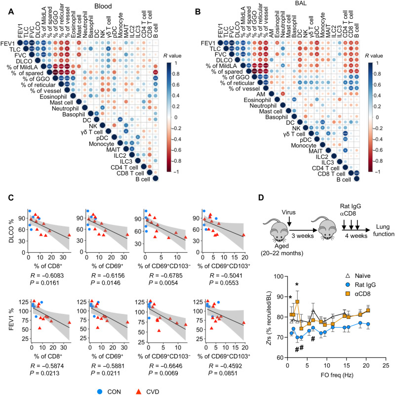
Severe coronavirus disease 2019 (COVID-19) pneumonia survivors often exhibit long-term pulmonary sequelae, but the underlying mechanisms or associated local and systemic immune correlates are not known. Here, we have performed high-dimensional characterization of the pathophysiological and immune traits of aged COVID-19 convalescents, and correlated the local and systemic immune profiles with pulmonary function and lung imaging. We found that chronic lung impairment was accompanied by persistent respiratory immune alterations. We showed that functional severe acute respiratory syndrome coronavirus 2 (SARS-CoV-2)–specific memory T and B cells were enriched at the site of infection compared with those of blood. Detailed evaluation of the lung immune compartment revealed that dysregulated respiratory CD8+ T cell responses were associated with the impaired lung function after acute COVID-19. Single-cell transcriptomic analysis identified the potential pathogenic subsets of respiratory CD8+ T cells contributing to persistent tissue conditions after COVID-19. Our results have revealed pathophysiological and immune traits that may support the development of lung sequelae after SARS-CoV-2 pneumonia in older individuals, with implications for the treatment of chronic COVID-19 symptoms.


Figures

Fig. 1.
Fig. 1.. Functional and pathological characterization of lung sequelae in aged COVID-19 convalescents.
(A) Schematics of experimental procedure. (B) Summary of control (CON) and COVID-19 convalescent age, sex, lung function, and condition data. (C) Lung regions of pathology revealed by quantitative CT analysis by CALIPER (green/dark green, “spared/relatively spared”; yellow, “GGO”; orange, “reticular/consolidation”). (D) Correlation of lung functional parameters with lung pathological features. Significant correlations were indicated by white asterisks. (E) Correlation of FEV1, FVC, and DLCO with % of mild lung area (MildLA), % of GGO region, and % of reticular regions in the lung. Significant correlations were indicated by asterisks. *P < 0.05.
Fig. 2.
Fig. 2.. Characterization of respiratory and circulating immune memory in COVID-19 convalescents.
(A) Percentage of indicated innate immune cell types in the whole blood (left) or adaptive immune cells in PBMCs (right). (B) Percentage of indicated immune cell types in the BAL. (C) Percentage of RBD-specific B cells within B cell compartment of BAL or PBMC. (D) CD69, CD27, IgD, and CD38 expression levels of RBD-specific B cells in BAL or PBMC. (E) Percentage of indicated CD8+ T cell subsets based on live cells in the BAL or PBMC. (F) Percentage of indicated CD8+ T cell subsets in total CD8+ T cells. (G) Percentage of IFN-γ, TNF, or interleukin (IL)–2+ CD8+ T cells in the BAL or PBMC after stimulation with SARS-CoV-2 peptide pools. (H) Percentage of indicated CD4+ T cell subsets based on live cells in the BAL or PBMC. (I) Percentage of IFN-γ, TNF, or IL-2+ CD4+ T cells in the BAL or PBMC after stimulation with SARS-CoV-2 peptide pools. (A, B, E, F, and H) Statistical significance was calculated using Mann-Whitney test. *P < 0.05. (C) Statistical difference was performed using two-way ANOVA. (D) Statistical significance was calculated using paired t test. (G and I) Statistical significance was calculated using two-way ANOVA following Fisher’s least significant difference (LSD) test. *P < 0.05.
Fig. 3.
Fig. 3.. Dysregulated CD8+ T cell responses as a potential driver of chronic lung sequelae.
(A) Correlation of blood immune cell types with lung functional parameters and quantitative lung pathology parameters. (B) Correlation of BAL immune cell types with lung functional parameters and quantitative lung pathology parameters. (C) Correlation of DLCO and FEV1 with % of total CD8+, CD69+ CD8+, CD69+CD103 CD8+, or CD69+CD103+ CD8+ T cells in the total BAL cells of healthy control and COVID-19 convalescents. (D) Aged mice were infected or not (naïve) with influenza virus. Mice receiving CD8+ T cell–depleting antibody (αCD8) or control antibody (rat IgG) starting at 21 days after infection. Lung function was measured at 50 days after infection. Input impedance (Zrs) was measured during tidal breathing and after recruitment of closed airways and displayed as the percent utilization at baseline for each frequency in the repeated forced oscillation (FO) waveform. (A and B) R values are indicated by color and circle size. Significant correlations were indicated by white asterisks. (D) Lung function data were cumulative from three independent experiments, and raw data can be found in fig. S9. *P < 0.05 for rat IgG (n = 16) versus CD8-depleted group (n = 8) or naïve (n = 7) versus rat IgG group (#) for two-way ANOVA following Fisher’s LSD test.
Fig. 4.
Fig. 4.. scRNA-seq analysis of respiratory CD8+ T cells from COVID-19 convalescents.
(A) BAL CD8+ T cell subclusters and relative abundance revealed by scRNA-seq. (B) Signature gene expression by different BAL CD8+ T cell clusters. (C) Relative levels of tissue residency, memory, and effector gene signatures in BAL CD8+ T cell clusters. (D) GSEA of autoaggressive CXCR6+ CD8+ T cells between clusters 0 and 2. (E) Different pathways enriched in cluster 2 compared with cluster 0 of BAL CD8+ T cells. (F) Relative abundance of BAL CD8+ T cell subclusters from control [aged-matched control recruited in this study and age-unmatched control from GSE151928 (42)] or COVID-19 convalescent donors. (G) Correlation of DLCO, FEV1, GGO, and reticular region with % of cluster 2 CXCR6hi CD8+ T cells in the BAL cells of age-matched control and COVID-19 convalescents.

References

    1. Polidori M. C., Sies H., Ferrucci L., Benzing T., COVID-19 mortality as a fingerprint of biological age. Ageing Res. Rev. 67, 101308 (2021). - PMC - PubMed
    1. Yang X., Yu Y., Xu J., Shu H., Xia J., Liu H., Wu Y., Zhang L., Yu Z., Fang M., Yu T., Wang Y., Pan S., Zou X., Yuan S., Shang Y., Clinical course and outcomes of critically ill patients with SARS-CoV-2 pneumonia in Wuhan, China: A single-centered, retrospective, observational study. Lancet Respir. Med. 8, –475, 481 (2020). - PMC - PubMed
    1. Wu X., Liu X., Zhou Y., Yu H., Li R., Zhan Q., Ni F., Fang S., Lu Y., Ding X., Liu H., Ewing R. M., Jones M. G., Hu Y., Nie H., Wang Y., 3-month, 6-month, 9-month, and 12-month respiratory outcomes in patients following COVID-19-related hospitalisation: A prospective study. Lancet Respir. Med. 9, 747–754 (2021). - PMC - PubMed
    1. Nalbandian A., Sehgal K., Gupta A., Madhavan M. V., McGroder C., Stevens J. S., Cook J. R., Nordvig A. S., Shalev D., Sehrawat T. S., Ahluwalia N., Bikdeli B., Dietz D., Der-Nigoghossian C., Liyanage-Don N., Rosner G. F., Bernstein E. J., Mohan S., Beckley A. A., Seres D. S., Choueiri T. K., Uriel N., Ausiello J. C., Accili D., Freedberg D. E., Baldwin M., Schwartz A., Brodie D., Garcia C. K., Elkind M. S. V., Connors J. M., Bilezikian J. P., Landry D. W., Wan E. Y., Post-acute COVID-19 syndrome. Nat. Med. 27, 601–615 (2021). - PMC - PubMed
    1. Xu Y. H., Dong J. H., An W. M., Lv X. Y., Yin X. P., Zhang J. Z., Dong L., Ma X., Zhang H. J., Gao B. L., Clinical and computed tomographic imaging features of novel coronavirus pneumonia caused by SARS-CoV-2. J. Infect. 80, 394–400 (2020). - PMC - PubMed

Publication types