. 2021 Dec 2;28(12):2104-2121.e10.
doi: 10.1016/j.stem.2021.09.002. Epub 2021 Sep 29.

Reprogramming reactive glia into interneurons reduces chronic seizure activity in a mouse model of mesial temporal lobe epilepsy

Affiliations

Reprogramming reactive glia into interneurons reduces chronic seizure activity in a mouse model of mesial temporal lobe epilepsy

Célia Lentini et al. Cell Stem Cell. .

Abstract

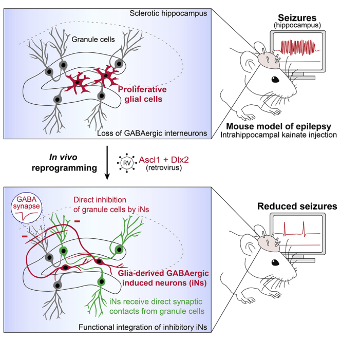
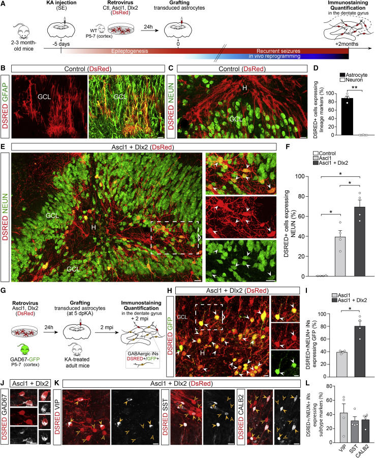
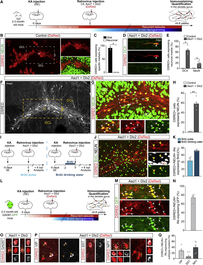
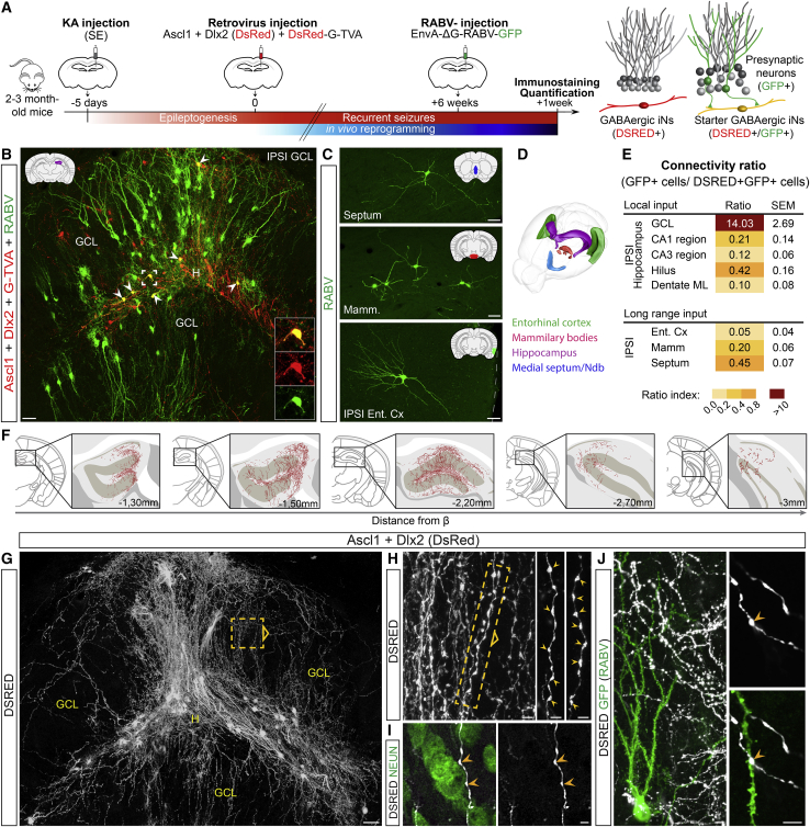
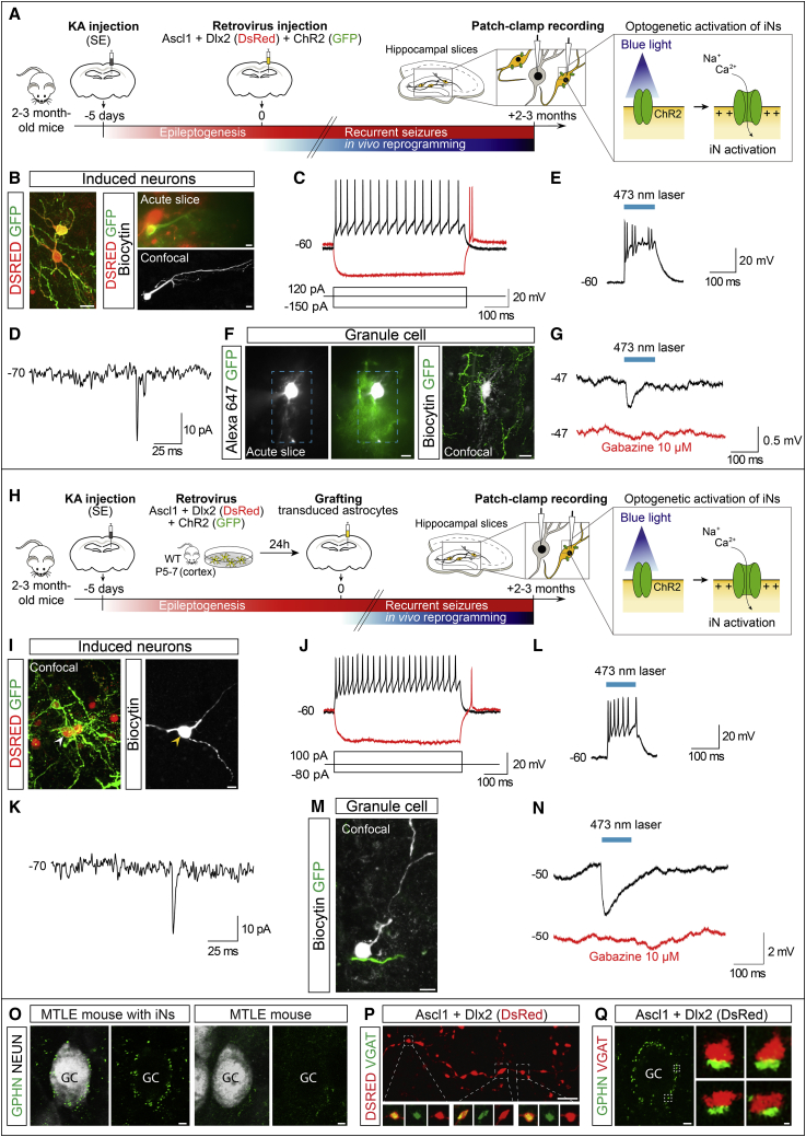
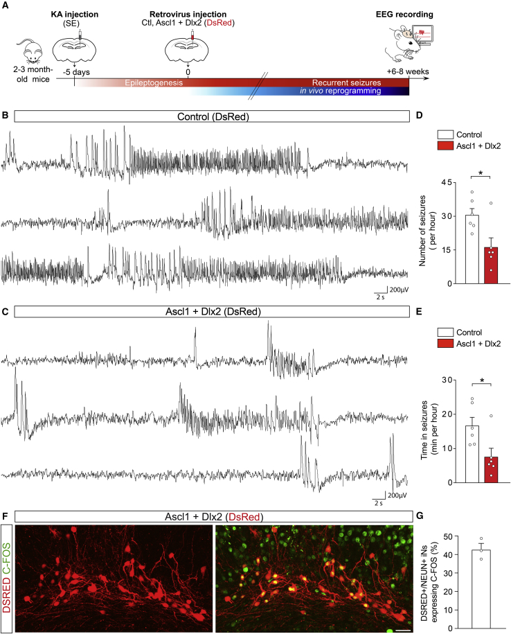
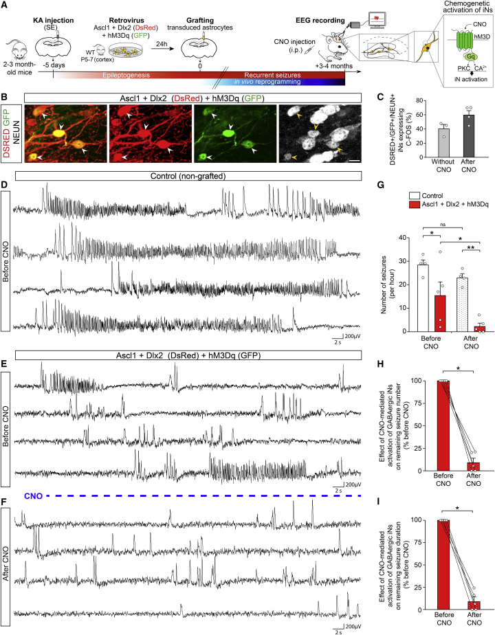
Reprogramming brain-resident glial cells into clinically relevant induced neurons (iNs) is an emerging strategy toward replacing lost neurons and restoring lost brain functions. A fundamental question is now whether iNs can promote functional recovery in pathological contexts. We addressed this question in the context of therapy-resistant mesial temporal lobe epilepsy (MTLE), which is associated with hippocampal seizures and degeneration of hippocampal GABAergic interneurons. Using a MTLE mouse model, we show that retrovirus-driven expression of Ascl1 and Dlx2 in reactive hippocampal glia in situ, or in cortical astroglia grafted in the epileptic hippocampus, causes efficient reprogramming into iNs exhibiting hallmarks of interneurons. These induced interneurons functionally integrate into epileptic networks and establish GABAergic synapses onto dentate granule cells. MTLE mice with GABAergic iNs show a significant reduction in both the number and cumulative duration of spontaneous recurrent hippocampal seizures. Thus glia-to-neuron reprogramming is a potential disease-modifying strategy to reduce seizures in therapy-resistant epilepsy.

Keywords: direct lineage reprogramming; gene therapy; glia-to-neuron conversion; regeneration and repair in the nervous system; regenerative medicine; therapy-resistant epilepsy.


Conflict of interest statement

Declaration of interests The authors declare no competing interests.

Figures

None
Graphical abstract
Figure 1
Figure 1
In vivo reprogramming of grafted cortical astroglia into GABAergic iNs within the MTLE-HS mouse hippocampus (A) Experimental procedures. (B and C) DSRED+ grafted astroglia transduced with the control retrovirus (DsRed) express GFAP, 2 mpi (B). None of the DSRED+ cells express NEUN (C). (D) Proportion of control-transduced astroglia (DSRED) expressing astrocytic (GFAP; n = 4) or neuronal markers (DCX, MAP2, or NEUN; n = 6), 2 mpi. (E) DSRED/NEUN+ iNs (arrowheads) derived from grafted astroglia transduced with the Ascl1/Dlx2-encoding retrovirus (DsRed), 2 mpi. Note the pronounced dispersion of dentate GCs induced by KA. (F) Proportion of DSRED+ cells converted into NEUN+ iNs following expression of Ascl1 (n = 4), Ascl1/Dlx2 (n = 4) or control (n = 4), 2 mpi. (G–I) Conversion of astroglia isolated from GAD67-GFP mice into GABAergic iNs. (G) Experimental procedures. (H) Ascl1/Dlx2-iNs (DSRED) expressing GFP (arrowheads; 2 mpi) demonstrating their GABAergic identity. (I) Proportion of DSRED/NEUN+ iNs expressing GFP following Ascl1 (n = 4) or Ascl1/Dlx2 (n = 4) reprogramming, 2 mpi. (J) Ascl1/Dlx2-iNs (DSRED) express GAD67. (K) Ascl1/Dlx2-iNs (DSRED) expressing VIP, SST, or CALB2 (full arrowheads). Empty arrowheads point to marker-negative iNs. (L) Proportion of DSRED/NEUN+ iNs expressing VIP, SST, or CALB2 (n = 4 each). Bars, mean ± SEM. Statistical analysis (D, F, and I): two-tailed Mann-Whitney test. p < 0.05, ∗∗p < 0.01. Right panels (E and H): magnified views of boxed areas. Composite images, (C) and (E). Scale bars: 25 μm (B, C, and E), 10 μm (H, J, and K). GCL, granule cell layer; H, hilus. See also Figures S1 and S2.
Figure 2
Figure 2
In vivo reprogramming of reactive hippocampal glia into GABAergic iNs in adult MTLE-HS mice (A) Experimental procedures. (B) DSRED+ hippocampal glia transduced with the control retrovirus (DsRed), 6 wpi. None of the DSRED+ cells express NEUN. (C) Proportion of control-transduced cells (DSRED) expressing glial (OLIG2, GFAP, or IBA1; n = 3) or neuronal marker (NEUN; n = 5), 6 wpi. (D–H) Hippocampal reactive glia transduced with the Ascl1/Dlx2-encoding retrovirus (DsRed) are reprogrammed into iNs. (D) DSRED+ iN expressing DCX, 7 dpi. (E) Proportion of DSRED+ cells expressing DCX or NEUN following expression of Ascl1/Dlx2 (DCX, n = 5; NEUN, n = 6) or control (n = 3), 7 dpi. (F) Ascl1/Dlx2-iNs (DSRED, white) exhibit complex neuronal morphologies and extend fibers creating dense networks throughout the dentate gyrus, 6 wpi. (G) Magnified views of the area boxed in (F) showing that Ascl1/Dlx2-iNs (DSRED) express NEUN (arrowheads), 6 wpi. (H) Proportion of DSRED+ cells converted into NEUN+ iNs following expression of Ascl1/Dlx2 (n = 6) or control (n = 5; same mice as in C), 6 wpi. (I–K) BrdU labeling of iNs. (I) Experimental procedures. (J) DSRED/NEUN+ iNs labeled by BrdU (arrowheads, drinking water protocol), 6 wpi. (K) Proportion of DSRED/NEUN+ iNs immunoreactive for BrdU following single BrdU pulse (n = 4) or BrdU supply in drinking water (n = 4), 6 wpi. (L–N) Conversion of hippocampal reactive glia into GABAergic iNs in GAD67-GFP mice. (L) Experimental procedures. (M) Ascl1/Dlx2-iNs (DSRED) expressing GFP (arrowheads; 6 wpi) demonstrating their GABAergic identity. (N) Proportion of DSRED/NEUN+ iNs expressing GFP following Ascl1/Dlx2 expression (n = 5), 6 wpi. (O) Ascl1/Dlx2-iNs (DSRED) express GAD67. (P) Ascl1/Dlx2-iNs (DSRED) expressing VIP, SST, or NPY (full arrowheads). Empty arrowhead points to a VIP-negative iN. (Q) Proportion of DSRED/NEUN+ iNs expressing VIP (n = 5), SST (n = 7), or NPY (n = 5), 8 wpi. Bars, mean ± SEM. Statistical analysis (C, E, and H): two-tailed Mann Whitney test. p < 0.05, ∗∗p < 0.01. Right (B and J) and bottom (G) panels: magnified views of boxed areas. Composite images, (B), (F), and (G). Scale bars: 10 μm except 25 μm (B and F). See also Figure S3.
Figure 3
Figure 3
iNs derived from hippocampal reactive glia show widespread synaptic integration within the MTLE-HS mouse brain (A) Experimental procedures. (B–E) iNs receive synaptic innervation from endogenous neurons, 7 wpi. (B) DSRED/GFP+ starter iNs (arrowheads, insets) receive innervation from local GFP+ GCs. (C) Starter iNs receive innervation from GFP+ long-range projection neurons in the ipsilateral entorhinal cortex (Ent Cx), mammillary/supramammillary bodies (Mamm), and medial septum/nucleus of diagonal band (Ndb). Schematics in (B) and (C) highlight the brain structure shown in each panel. (D) 3D drawing shows brain regions establishing synapses onto iNs. (E) Numbers of GFP+ presynaptic neurons expressed as color-coded connectivity ratios, 7 wpi (mean ± SEM; n = 3). (F–J) iNs send axons impinging on GCs, 7 wpi (n = 8). (F) DSRED+ fibers from iNs extend over 1.7 mm along the rostro-caudal axis of the dorsal hippocampus. (G) Ascl1/Dlx2-iNs (DSRED, white) extend fibers forming dense networks throughout the dentate gyrus. (H) Magnified view of the area boxed in (G) shows iN axons and synaptic bouton-like structures (arrowheads). Right panels: magnified views of boxed area. (I) Synaptic boutons in close contact (arrowheads) with a NEUN+ GC soma. (J) iNs extend axons across dendritic arbors of GCs (GFP from RABV as in B). Synaptic boutons contact GC dendrites. Composite images, (B) and (G). Scale bars: 50 μm (B, C, and G), 5 μm (H–J). ML, molecular layer. See also Figure S4.
Figure 4
Figure 4
iNs are physiologically functional and form GABAergic synapses with GCs in the MTLE-HS hippocampus (A–G) iNs derived from hippocampal reactive glia. (A) Experimental procedures. (B) Left: Ascl1/Dlx2-iNs (DSRED) expressing CHR2 (GFP). Right: example of recorded DSRED/CHR2+ iN visualized in acute slice (top), and after recording and intracellular biocytin injection (bottom). (C) iN showing repetitive action potential firing in response to depolarizing current injection (black; 4 iNs recorded, n = 3 mice). Rebound spiking generated following relief from hyperpolarization (red; 3 of 4 iNs). (D) Spontaneous synaptic input recorded from an iN (3 of 3 iNs, n = 3 mice). (E) DSRED/CHR2+ iN showing action potential firing in response to blue light stimulation (473 nm, 100 ms; 4 of 4 iNs, n = 3 mice). (F) Example of recorded GC (filled with Alexa 647) surrounded by CHR2+ (GFP) iN processes visualized in acute slice (left), and after recording and intracellular biocytin injection (right). Blue box: area of laser stimulation. (G) GABAergic IPSPs recorded in a GC (average trace of 5 consecutive responses is shown) evoked by blue light stimulation of CHR2+ iNs (black; 4 of 8 GCs, n = 3 mice). IPSPs were blocked by gabazine (red; 4 of 4 GCs, n = 3 mice). (H–N) iNs derived from grafted cortical glia. (H) Experimental procedures. (I) Recorded DSRED/CHR2+ iN filled with biocytin (arrowhead). (J) Repetitive action potential firing (black; 4 iNs recorded, n = 4 mice) and rebound spike (red; 3 of 4 iNs) recorded in an iN as in (C). (K) iNs receive spontaneous synaptic input (3 of 4 iNs, n = 4 mice). (L) iN activation by blue light (4 of 4 iNs, n = 4 mice). (M) Recorded GC (filled with biocytin) surrounded by a CHR2+ iN process. (N) GABAergic IPSPs recorded in a GC as in (G) (black; 6 of 8 GCs, n = 5 mice) and blocked by gabazine (red, 4 of 4 GCs, n = 4 mice). (O–Q) Immunohistological evidence for GABAergic synapses between iNs and GCs. (O) Left: example of a GC (white) exhibiting gephyrin+ (GPHN) puncta outlining its soma in MTLE-HS mice after Ascl1/Dlx2-reprogramming, 2 mpi. Right: very few gephyrin+ puncta in MTLE-HS mice without iNs. (P) DSRED+ axon from a GABAergic iN showing axonal varicosities containing clusters of VGAT+ puncta. (Q) Gephyrin+ puncta around a GC soma (left) and examples of close apposition of gephyrin+ and VGAT+ puncta (right) in mice with GABAergic iNs. Scale bars: 10 μm (B, F, I, and M), 2 μm (O–Q). See also Figure S5.
Figure 5
Figure 5
In vivo reprogramming of hippocampal reactive glia into GABAergic iNs reduces spontaneous recurrent hippocampal seizures in MTLE-HS mice (A) Experimental procedures. (B) Intrahippocampal EEG recording from a MTLE-HS mouse injected with the control retrovirus (DsRed) show numerous non-convulsive EEG seizures consisting of slow rhythmic high-voltage sharp waves followed by higher-frequency and lower-amplitude spikes (6–8 wpi). (C) Intrahippocampal EEG recording from a MTLE-HS mouse injected with the Ascl1/Dlx2-retrovirus show drastic decrease in number of hippocampal EEG seizures and time spent in seizures (6–8 wpi). (D and E) Number of hippocampal EEG seizures (D) and time spent in seizures (E, cumulative seizure duration, min/h) in MTLE-HS mice injected with the Ascl1/Dlx2-retrovirus (n = 6) or control retrovirus (n = 6), 6–8 wpi. Statistical analysis: two-tailed Mann-Whitney test. p < 0.05. (F and G) A substantial fraction of iNs is physiologically active. (F) Numerous Ascl1/Dlx2-iNs (DSRED) express C-FOS, 8 wpi. (G) Proportion of DSRED/NEUN+ iNs immunoreactive for C-FOS, 6–8 wpi (n = 3). Representative traces in (B) and (C) show EEG recordings (3 min each) in the KA-injected hippocampus. Bars, mean ± SEM. Scale bar: 25 μm. See also Figure S6.
Figure 6
Figure 6
Chemogenetically enhancing iN activity results in complete seizure suppression in MTLE-HS mice (A) Experimental procedures. (B) NEUN+ (white) iNs derived from grafted astroglia expressing Ascl1/Dlx2 (DSRED) and hM3Dq (GFP, arrowheads), 4 mpi. (C) Proportion of DSRED/NEUN/GFP+ iNs immunoreactive for C-FOS in absence of CNO (n = 3) and after CNO treatment (n = 4), 3–4 mpi. (D) Intrahippocampal EEG recording from a control non-grafted MTLE-HS mouse showing recurrence of numerous EEG seizures. (E and F) Intrahippocampal EEG recordings from a MTLE-HS mouse with hM3Dq+ GABAergic iNs before (E) and after CNO treatment (F), 3–4 mpi. (E) Drastic decrease in the number of EEG seizures in absence of CNO compared to control non-grafted MTLE-HS mice (D). (F) CNO-evoked activation of hM3Dq+ iNs suppresses remaining EEG seizure activity below reducing effects of iNs in absence of CNO (E). Only residual isolated spikes remain visible. (G) Number of hippocampal EEG seizures in MTLE-HS mice with hM3Dq+ GABAergic iNs (red bars; n = 5) and control non-grafted MTLE-HS mice (white bars; n = 4) in absence of CNO (left bars) and after CNO treatment (right dotted bars). Same mice analyzed before and after CNO. Statistical analysis: repeated-measures two-way ANOVA followed by Sidak’s multiple comparison post hoc test. p < 0.05, ∗∗p < 0.01. (H and I) Pairwise comparisons of seizure numbers (H) and time in seizures (I) before and after CNO treatment in each MTLE-HS mouse with hM3Dq+ GABAergic iNs (n = 5; same mice as in G red bars). Statistical analysis: one-tailed Wilcoxon matched-pairs test. p < 0.05. Lines connect data from individual mice before and after CNO. Representative traces in (D)–(F) show EEG recordings (4 min each) in the KA-injected hippocampus. Bars, mean ± SEM. Scale bar: 10 μm. See also Figure S6.

Comment in

  • In the hands of fate change.
    Lewis S. Lewis S. Nat Rev Neurosci. 2021 Dec;22(12):720-721. doi: 10.1038/s41583-021-00537-6. Nat Rev Neurosci. 2021. PMID: 34703022 No abstract available.

References

    1. Alvarez D.D., Giacomini D., Yang S.M., Trinchero M.F., Temprana S.G., Büttner K.A., Beltramone N., Schinder A.F. A disynaptic feedback network activated by experience promotes the integration of new granule cells. Science. 2016;354:459–465. - PubMed
    1. Andrioli A., Alonso-Nanclares L., Arellano J.I., DeFelipe J. Quantitative analysis of parvalbumin-immunoreactive cells in the human epileptic hippocampus. Neuroscience. 2007;149:131–143. - PubMed
    1. Arabadzisz D., Antal K., Parpan F., Emri Z., Fritschy J.M. Epileptogenesis and chronic seizures in a mouse model of temporal lobe epilepsy are associated with distinct EEG patterns and selective neurochemical alterations in the contralateral hippocampus. Exp. Neurol. 2005;194:76–90. - PubMed
    1. Baraban S.C., Southwell D.G., Estrada R.C., Jones D.L., Sebe J.Y., Alfaro-Cervello C., García-Verdugo J.M., Rubenstein J.L., Alvarez-Buylla A. Reduction of seizures by transplantation of cortical GABAergic interneuron precursors into Kv1.1 mutant mice. Proc. Natl. Acad. Sci. USA. 2009;106:15472–15477. - PMC - PubMed
    1. Barker R.A., Götz M., Parmar M. New approaches for brain repair-from rescue to reprogramming. Nature. 2018;557:329–334. - PubMed

Publication types

LinkOut - more resources