. 2021 Sep 30;11(1):19446.
doi: 10.1038/s41598-021-98102-9.

Vm-related extracellular potentials observed in red blood cells

Affiliations

Vm-related extracellular potentials observed in red blood cells

Michael Pycraft Hughes et al. Sci Rep. .

Abstract

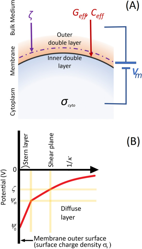
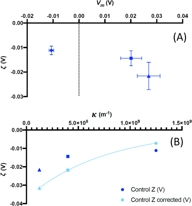
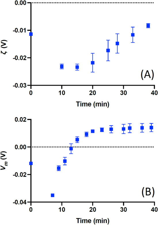
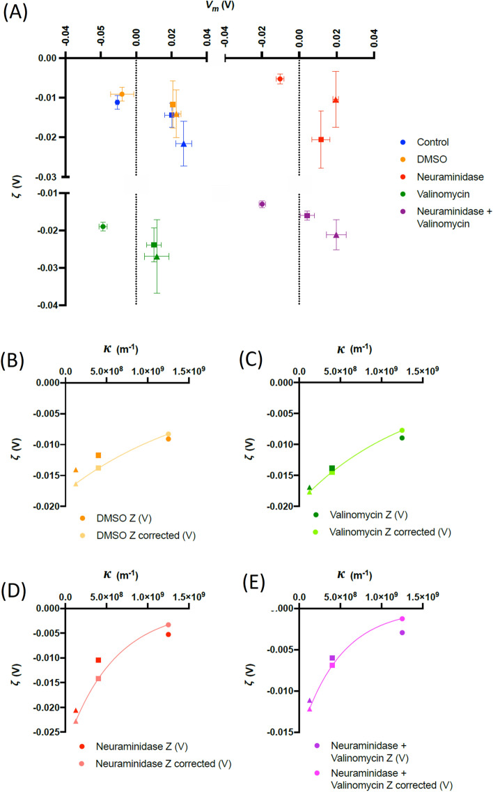
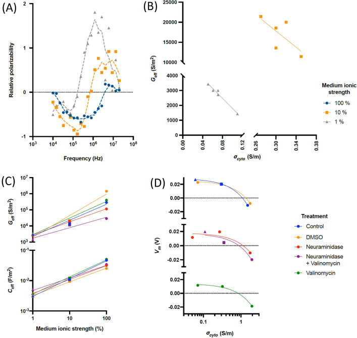
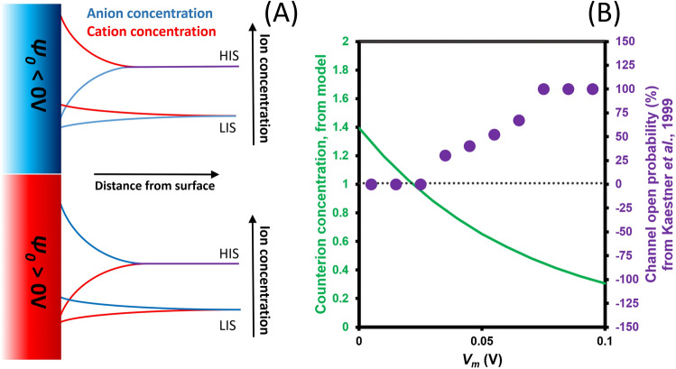
Even in nonexcitable cells, the membrane potential Vm is fundamental to cell function, with roles from ion channel regulation, development, to cancer metastasis. Vm arises from transmembrane ion concentration gradients; standard models assume homogeneous extracellular and intracellular ion concentrations, and that Vm only exists across the cell membrane and has no significance beyond it. Using red blood cells, we show that this is incorrect, or at least incomplete; Vm is detectable beyond the cell surface, and modulating Vm produces quantifiable and consistent changes in extracellular potential. Evidence strongly suggests this is due to capacitive coupling between Vm and the electrical double layer, rather than molecular transporters. We show that modulating Vm changes the extracellular ion composition, mimicking the behaviour if voltage-gated ion channels in non-excitable channels. We also observed Vm-synchronised circadian rhythms in extracellular potential, with significant implications for cell-cell interactions and cardiovascular disease.


Conflict of interest statement

MPH is a director of Deparator, which manufactures the 3DEP instrument used for the DEP measurements. The authors declare no other interests.

Figures

Figure 1
Figure 1
(A) A schematic of the cell, indicating the location of the electrical double layers, the shear plane (broken purple line) of ζ, the specific membrane conductance Geff and capacitance Ceff, cytoplasm conductivity σcyto and membrane potential Vm. (B) The variation in potential from the cell surface, showing the location of the Stern layer and the shear plane at which ζ is located.
Figure 2
Figure 2
(A) The mean variation (± s.d.) in Vm and ζ for control RBCs in media containing 100% (square), 10% (circle) and 1% (triangle) physiological saline as described in the text. (B) The same values of ζ plotted against the calculated reciprocal Debye length κ for each medium, together with values adjusted by 0.37 Vm. The relationship between these variables theoretically follows a negative exponential to which the adjusted values offer a perfect (r2 = 1) fit. All points represent the average of four donors.
Figure 3
Figure 3
The mean (n = 3) Vm (A) and ζ-potential (B) of RMCE immediately after resuspension into 10% solution (see text). As can be seen, in both instances the value immediately changes from a rest value (taken in 100% solution, denoted here as time 0), peaks and then increases to a rest value. The transition period was consistent for both Vm and ζ, but the former was observed to respond consistently across all three samples, whereas for ζ a delay of 5–15 min was observed before transition began.
Figure 4
Figure 4
(A) The mean variation (± s.d.) in Vm and ζ for RBCs suspended in media containing 100% (square), 10% (circle) and 1% (triangle) physiological saline as described in the text. RBCs were treated with valinomycin, neuraminidase, and a combination of both, as well as a DMSO was control. All points represent the average of four donors. (BE) The values of ζ from Fig. 4A plotted against the reciprocal Debye length κ, together with values adjusted by ΞVm as described in the text. The relationship between these variables theoretically follows a negative exponential, to which the adjusted values offer a perfect (R2 = 1) fit. All points represent the average of four donors.
Figure 5
Figure 5
(A) The DEP spectra (points) of RBCs in media of different ionic strength (1% (grey), 10% (orange) and 100% (blue)) together with rolling average trendlines; (B) the values of Geff vs. σcyto for 1% and 10% media, together with best-fit trendline; (C) Specific membrane conductance Geff and capacitance Ceff as a function of medium ionic strength; (D) the relationship between cytoplasm conductivity σcyto and membrane potential Vm.
Figure 6
Figure 6
Circadian behaviour of ζ-potential in RBCs. (A) mean (n = 4) ζ for RBCs entrained and suspended in KHB and DEP medium, together with best-fit rhythm with period of 24.5 h and 24.3 h respectively. (B) ζ of RBCs taken directly from a participant over a 24 h period, measured within 60 s of donation, together with a best-fit rhythm with 22.2 h period (time 0 h corresponds to 10am).
Figure 7
Figure 7
(A) A schematic showing ion concentrations in proximity to the cell surface in high ionic strength (HIS) and low ionic strength (LIS) solutions. Where the surface potential of a cell is negative, it will electrostatically attract cations from solution, raising the cation concentration at the surface whilst depleting anions. Where the surface potential is positive, this situation is reversed, with the cation level in HIS solutions reaching similar values to those observed in LIS solutions. (B) A comparison of the voltage-gated cation channel behaviour reported by Kaestner et al., and the cation concentration at the surface relative to the bulk (green line) determined using the Poisson-Boltzmann equation, using ζ determined from Eq. (5).

References

    1. Galvani, L. De viribus electricitatis in motu musculari commentarius (Ex Typographia Instituti Scientiarum; 1792).
    1. Nernst, W.H. Die elektromotorische wirksamkeit der Jonen (W. Engelmann, 1889).
    1. Goldman DE. Potential, impedance, and rectification in membranes. J. Gen. Physiol. 1943;27:37–60. doi: 10.1085/jgp.27.1.37. - DOI - PMC - PubMed
    1. Hodgkin AL, Katz B. The effect of sodium ions on the electrical activity of the giant axon of the squid. J. Physiol. 1949;108:37–77. doi: 10.1113/jphysiol.1949.sp004310. - DOI - PMC - PubMed
    1. Hodgkin AL, Huxley AF. A quantitative description of membrane current and its application to conduction and excitation in nerve. J. Physiol. 1952;117:500–544. doi: 10.1113/jphysiol.1952.sp004764. - DOI - PMC - PubMed