. 2021 Sep 30;11(1):19523.
doi: 10.1038/s41598-021-98588-3.

A novel thermostable prokaryotic fucoidan active sulfatase PsFucS1 with an unusual quaternary hexameric structure

Affiliations

A novel thermostable prokaryotic fucoidan active sulfatase PsFucS1 with an unusual quaternary hexameric structure

Maria Dalgaard Mikkelsen et al. Sci Rep. .

Abstract

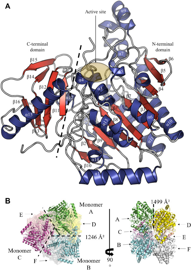
Fucoidans are sulfated, fucose-rich marine polysaccharides primarily found in cell walls of brown seaweeds (macroalgae). Fucoidans are known to possess beneficial bioactivities depending on their structure and sulfation degree. Here, we report the first functional characterization and the first crystal structure of a prokaryotic sulfatase, PsFucS1, belonging to sulfatase subfamily S1_13, able to release sulfate from fucoidan oligosaccharides. PsFucS1 was identified in the genome of a Pseudoalteromonas sp. isolated from sea cucumber gut. PsFucS1 (57 kDa) is Ca2+ dependent and has an unusually high optimal temperature (68 °C) and thermostability. Further, the PsFucS1 displays a unique quaternary hexameric structure comprising a tight trimeric dimer complex. The structural data imply that this hexamer formation results from an uncommon interaction of each PsFucS1 monomer that is oriented perpendicular to the common dimer interface (~ 1500 Å2) that can be found in analogous sulfatases. The uncommon interaction involves interfacing (1246 Å2) through a bundle of α-helices in the N-terminal domain to form a trimeric ring structure. The high thermostability may be related to this unusual quaternary hexameric structure formation that is suggested to represent a novel protein thermostabilization mechanism.


Conflict of interest statement

The authors declare no competing interests.

Figures

Figure 1
Figure 1
PsFucS1 fold and oligomeric state. (A) Cartoon representation of the PsFucS1 structure showing the domain organization and secondary structure elements. The N-terminal domain (Asn21-Leu419: α1-α14 helices and β1–β10 strands) and the C-terminal domain (Val430-Gly521: α15 helix and β11–β16 strands) are separated by a dashed line. The active site position is circled in yellow. Secondary structure elements are colored in red and blue for β-strands and α-helices, respectively. (B) Cartoon representation of the PsFucS1 hexamer (trimer of dimers) in the asymmetric unit. Monomers A, B, C, D, E and F are colored in green, cyan, purple, yellow, pink and white, respectively.
Figure 2
Figure 2
PsFucS1 active site topology. (A,B) Cartoon representation of PsFucS1. The N-terminal domain and the C-terminal domain are colored in blue and red, respectively. The active site pocket is circled in yellow. Structural elements delineating the protein active site are indicated by arrows. In (B) Part of the interfacing neighboring monomer is indicated in green including the α15 helix that partially covers the active site. (C) PsFucS1 crystal structure in electrostatic representation. The active site pocket is circled in yellow. (D) Electrostatic representation of the endo-4S-ι-carrageenan sulfatase from Pseudoalteromonas sp. PS47 (6B0J) in complex with κ-ι-κ-neocarrahexaose shown for comparison. The active site groove is circled in yellow. Electrostatic potential is expressed as a spectrum ranging from − 5 kT e−1 (red) to + 5 kT e−1 (blue) and was calculated in APBS. (E) Electron density around the calcium ion of PsFucS1. The map shown is a σA-weighted 2mFo-DFc map contoured at 1.2σ (0.67e Å−3). Asp69, Asp70, Cys111, Asp334 and Asn335 residues are shown as sticks. The calcium atom is shown as a green sphere.
Figure 3
Figure 3
Docking results of sugar-sulfatase complexes and PsFucS1 activity on fucoidan. (A) Active site pocket of PsFucS1 X-ray crystal structure. (B) Cartoon representation of C2 sulfated fucobiose-PsFucS1 complex obtained by docking. The red S indicates the position of the sulfate binding site as defined by Hettle et al. 2018. (C) Zoomed-in view of PsFucS1 active site with docked disaccharide at the dimer interface. (AC) Monomers A and B are colored in green and cyan, respectively. Residues constituting the binding pocket are pointed as sticks and those from monomer A are labeled in green. Secondary structure elements are shown as cartoon. The calcium atom and water molecules are shown as green and red spheres, respectively. The bound disaccharide into the active site pocket is shown as purple sticks. H bonds between the ligand and protein are yellow dotted lines. (D) Superposition of the PsFucS1 X-ray crystal structure (white) to the monomer docked model (red) and the dimer docked model (blue).

References

    1. Bilan MI, et al. Structure of a fucoidan from the brown seaweed Fucus evanescens C. Ag. Carbohydr. Res. 2002;37:719–730. doi: 10.1016/S0008-6215(02)00053-8. - DOI - PubMed
    1. Thinh PD, et al. Structural characteristics and anticancer activity of fucoidan from the brown alga Sargassum mcclurei. Mar. Drugs. 2013;11:1456–1476. doi: 10.3390/md11051456. - DOI - PMC - PubMed
    1. Ale MT, Mikkelsen JD, Meyer AS. Important determinants for fucoidan bioactivity: A critical review of structure-function relations and extraction methods for fucose-containing sulfated polysaccharides from brown seaweeds. Mar. Drugs. 2011;9:2106–2130. doi: 10.3390/md9102106. - DOI - PMC - PubMed
    1. Torres MD, et al. Fucoidans: The importance of processing on their anti-tumoral properties. Algal Res. 2020;45:101748. doi: 10.1016/j.algal.2019.101748. - DOI
    1. Silchenko AS, et al. Structure, enzymatic transformation, anticancer activity of fucoidan and sulphated fucooligosaccharides from Sargassum horneri. Carbohydr. Polym. 2017;175:654–660. doi: 10.1016/j.carbpol.2017.08.043. - DOI - PubMed

Publication types