. 2021 Nov;22(5):1271.
doi: 10.3892/etm.2021.10706. Epub 2021 Sep 7.

TFEC contributes to cardiac hypertrophy by inhibiting AMPK/mTOR signaling

Affiliations

TFEC contributes to cardiac hypertrophy by inhibiting AMPK/mTOR signaling

Ting Zhao et al. Exp Ther Med. 2021 Nov.

Abstract

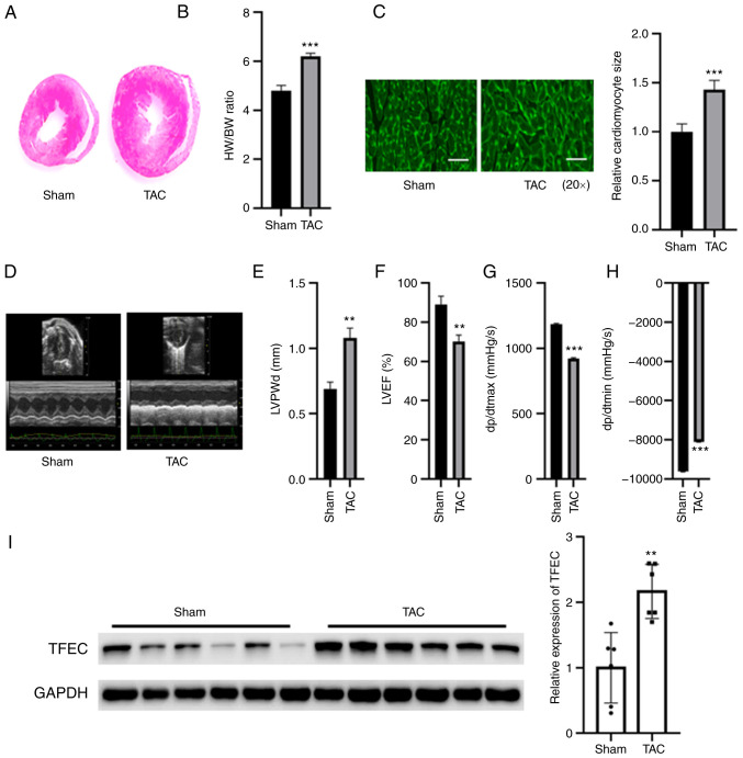
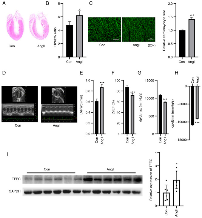
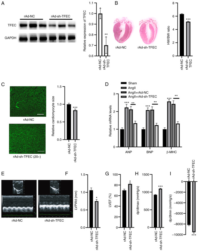
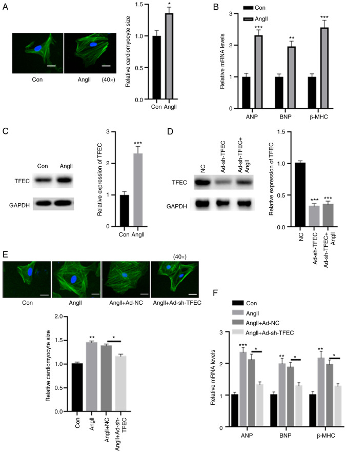
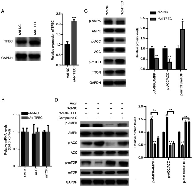
The underlying mechanism of cardiac hypertrophy has not yet been fully elucidated. The present study aimed to explore the function of transcription factor EC (TFEC) in mouse models of cardiac hypertrophy and to determine the underlying mechanism. Pressure-overload cardiac hypertrophy and angiotensin II (AngII) infusion-induced animal models of cardiac hypertrophy were established in vivo. The expression of TFEC was explored via western blotting. The results demonstrated that TFEC expression was significantly increased in the hearts of mice with pressure overload- and AngII-induced hypertrophy. Injection of rAd-short hairpin (sh)-TFEC significantly decreased the expression of TFEC in heart tissues compared with group injected with rAd-negative control (NC). Furthermore, the expression levels of atrial natriuretic peptide (ANP), brain natriuretic peptide (BNP) and β-myosin heavy chain (β-MHC) were increased in the hearts of AngII-treated mice; however, compared with rAd-NC transfection, transfection with rAd-sh-TFEC decreased the expression levels of ANP, BNP and β-MHC. The results from echocardiographic analysis indicated that transfection with rAd-sh-TFEC improved the cardiac function of AngII-treated mice compared with transfection with rAd-NC. In addition, the AngII-induced increase in cardiomyocyte size could be reversed by TFEC knockdown in primary cardiomyocytes. The elevated expression levels of ANP, BNP and β-MHC induced by AngII could be partially abolished following TFEC knockdown. The results from western blotting demonstrated that TFEC overexpression decreased the expression of phosphorylated AMP-activated protein kinase (AMPK)/acetyl-CoA carboxylase (ACC) but increased the expression of phosphorylated mechanistic target of rapamycin (mTOR). Furthermore, Compound C significantly suppressed the activation of AMPK/ACC but increased the activation of mTOR, even in primary cardiomyocytes transfected with rAd-sh-TFEC. In conclusion, the findings from this study demonstrated that TFEC was overexpressed in the hearts of mice with cardiac hypertrophy and that silencing TFEC may improve AngII-induced cardiac hypertrophy and dysfunction by activating AMPK/mTOR signaling.

Keywords: AMP-activated protein kinase/mechanistic target of rapamycin signaling; cardiac hypertrophy; kinase non-catalytic C-lobe domain containing 1.


Conflict of interest statement

The authors declare that they have no competing interests.

Figures

Figure 1
Figure 1
TFEC expression is increased in the hypertrophic myocardia of mice subjected to TAC. (A) Hematoxylin and eosin staining of heart tissues. (B) HW/BW was evaluated in the sham and TAC groups. (C) Wheat germ agglutinin staining showed that the cardiomyocyte size was significantly increased in the hypertrophic myocardia of mice subjected to TAC compared with control mice (magnification, x20). (D) Representative echocardiographic images. (E) LVPWd, (F) LVEF (%), (G) dp/dtmax and (H) dp/dtmin were quantified in the hearts of TAC and sham control groups. (I) Western blotting showed that TFEC expression was increased in the hypertrophic myocardia of mice subjected to TAC compared with control mice. **P<0.01 and ***P<0.001. TAC, transverse aortic constriction; TFEC, transcription factor EC; HW/BW, heart weight and body weight ratio; LVEF, left ventricular ejection fractions; LVPWd, left ventricular posterior wall diameter; dp/dtmax, maximum change in left ventricular pressure over time; dp/dtmin, minimum change in left ventricular pressure over time.
Figure 2
Figure 2
TFEC expression is elevated in the hearts of AngII-treated mice compared with control mice. (A) Hematoxylin and eosin staining of heart tissues. (B) HW/BW was evaluated in control and Ang-treated mice. (C) Wheat germ agglutinin staining was performed to evaluate the relative cardiomyocyte size (magnification, x20). (D) Representative echocardiographic images. (E) LVPWd, (F) LVEF (%), (G) dp/dtmax and (H) dp/dtmin were quantified in the hearts of AngII-treated mice and sham control mice. (I) Western blotting assay demonstrated that TFEC expression was increased in the hearts of AngII-treated mice compared with those of control mice. *P<0.05 and ***P<0.001. AngII, angiotensin II; TFEC, transcription factor EC; HW/BW, heart weight and body weight ratio; LVEF, left ventricular ejection fractions; LVPWd, left ventricular posterior wall diameter; dp/dtmax, maximum change in left ventricular pressure over time; dp/dtmin, minimum change in left ventricular pressure over time.
Figure 3
Figure 3
TFEC knockdown improves cardiac function in AngII-treated mice compared with NC mice. (A) Western blotting analysis showed that injection with rAd-sh-TFEC significantly decreased the expression of TFEC in heart tissues compared with injection with rAd-NC. (B) Knockdown of TFEC reduced the HW/BW compared with NC group. (C) Relative cardiomyocyte size was decreased in mice injected with rAd-sh-TFEC compared with those injected with rAd-NC (magnification, x20). (D) Transfection with rAd-sh-TFEC reduced the relative mRNA levels of ANP, BNP and β-MHC compared with transfection with rAd-NC. (E) Representative echocardiographic images. (F) LVPWd, (G) LVEF (%), (H) dp/dtmax and (I) dp/dtmin were quantified in the hearts of rAd-sh-TFEC and rAd-NC-treated mice. *P<0.05, **P<0.01 and ***P<0.001. TFEC, transcription factor EC; sh, short hairpin; NC, negative control; HW/BW, heart weight and body weight ratio; ANP, atrial natriuretic peptide; BNP, brain natriuretic peptide; β-MHC, β-myosin heavy chain; LVEF, left ventricular ejection fractions; LVPWd, left ventricular posterior wall diameter; dp/dtmax, maximum change in left ventricular pressure over time; dp/dtmin, minimum change in left ventricular pressure over time.
Figure 4
Figure 4
Silencing TFEC abolishes AngII-induced cardiomyocyte hypertrophy. (A) Compared with the control, treatment with AngII increased the relative cardiomyocyte size (magnification, x40). (B) mRNA levels of ANP, BNP and β-MHC were significantly increased in primary cardiomyocytes treated with AngII compared with the control. (C) In primary cardiomyocytes, treatment with AngII significantly increased the expression of TFEC compared with treatment with the control. (D) Transfection with rAd-sh-TFEC significantly decreased the expression of TFEC even in primary cardiomyocytes treated with AngII. (E) AngII-induced increase in cardiomyocyte size could be reversed by TFEC knockdown in primary cardiomyocytes (magnification, x40). (F) Elevated mRNA levels of ANP, BNP and β-MHC induced by AngII could be partially abolished following TFEC knockdown in primary cardiomyocytes. *P<0.05, **P<0.01 and ***P<0.001. AngII, angiotensin II; Con, control; NC, negative control; ANP, atrial natriuretic peptide; BNP, brain natriuretic peptide; β-MHC, β-myosin heavy chain; TFEC, transcription factor EC; sh, short hairpin.
Figure 5
Figure 5
TFEC activates AMPK/mTOR signaling in primary cardiomyocytes. (A) Western blotting showed that TFEC expression was significantly increased in primary cardiomyocytes transfected with Ad-TFEC compared with those transfected with Ad-NC. (B) Reverse transcription quantitative PCR showed that transfection with rAd-TFEC did not change the mRNA levels of AMPK, ACC and mTOR, compared with transfection with rAd-NC. (C) Western blotting showed that TFEC overexpression decreased the expression of p-AMPK and p-ACC but increased the level of p-mTOR. (D) Compound C significantly suppressed the activation of AMPK/ACC but increased the activation of mTOR, even in primary cardiomyocytes transfected with rAd-sh-TFEC. *P<0.05, **P<0.01 and ***P<0.001. AMPK, AMP-activated protein kinase; mTOR, mechanistic target of rapamycin; ACC, acetyl-CoA carboxylase; NC, negative control; p, phosphorylated; sh, short hairpin; TFEC, transcription factor EC.

References

    1. Nakamura M, Sadoshima J. Mechanisms of physiological and pathological cardiac hypertrophy. Nat Rev Cardiol. 2018;15:387–407. doi: 10.1038/s41569-018-0007-y. - DOI - PubMed
    1. Gallo S, Vitacolonna A, Bonzano A, Comoglio P, Crepaldi T. ERK: A key player in the pathophysiology of cardiac hypertrophy. Int J Mol Sci. 2019;20(2164) doi: 10.3390/ijms20092164. - DOI - PMC - PubMed
    1. Meng R, Pei Z, Zhang A, Zhou Y, Cai X, Chen B, Liu G, Mai W, Wei J, Dong Y. AMPK activation enhances PPARα activity to inhibit cardiac hypertrophy via ERK1/2 MAPK signaling pathway. Arch Biochem Biophys. 2011;511:1–7. doi: 10.1016/j.abb.2011.04.010. - DOI - PubMed
    1. Petratou K, Spencer SA, Kelsh RN, Lister JA. The MITF paralog tfec is required in neural crest development for fate specification of the iridophore lineage from a multipotent pigment cell progenitor. PLoS One. 2021;16(e0244794) doi: 10.1371/journal.pone.0244794. - DOI - PMC - PubMed
    1. Ballesteros-Alvarez J, Dilshat R, Fock V, Möller K, Karl L, Larue L, Ögmundsdóttir MH, Steingrímsson E. MITF and TFEB cross-regulation in melanoma cells. PLoS One. 2020;15(e0238546) doi: 10.1371/journal.pone.0238546. - DOI - PMC - PubMed