. 2021 Oct;10(12):e12152.
doi: 10.1002/jev2.12152.

Extracellular vesicles from adipose tissue-derived stem cells alleviate osteoporosis through osteoprotegerin and miR-21-5p

Affiliations

Extracellular vesicles from adipose tissue-derived stem cells alleviate osteoporosis through osteoprotegerin and miR-21-5p

Kyoung Soo Lee et al. J Extracell Vesicles. 2021 Oct.

Abstract

Osteoporosis is one of the most common skeletal disorders caused by the imbalance between bone formation and resorption, resulting in quantitative loss of bone tissue. Since stem cell-derived extracellular vesicles (EVs) are growing attention as novel cell-free therapeutics that have advantages over parental stem cells, the therapeutic effects of EVs from adipose tissue-derived stem cells (ASC-EVs) on osteoporosis pathogenesis were investigated. ASC-EVs were isolated by a multi-filtration system based on the tangential flow filtration (TFF) system and characterized using transmission electron microscopy, dynamic light scattering, zeta potential, flow cytometry, cytokine arrays, and enzyme-linked immunosorbent assay. EVs are rich in growth factors and cytokines related to bone metabolism and mesenchymal stem cell (MSC) migration. In particular, osteoprotegerin (OPG), a natural inhibitor of receptor activator of nuclear factor-κB ligand (RANKL), was highly enriched in ASC-EVs. We found that the intravenous administration of ASC-EVs attenuated bone loss in osteoporosis mice. Also, ASC-EVs significantly inhibited osteoclast differentiation of macrophages and promoted the migration of bone marrow-derived MSCs (BM-MSCs). However, OPG-depleted ASC-EVs did not show anti-osteoclastogenesis effects, demonstrating that OPG is critical for the therapeutic effects of ASC-EVs. Additionally, small RNA sequencing data were analysed to identify miRNA candidates related to anti-osteoporosis effects. miR-21-5p in ASC-EVs inhibited osteoclast differentiation through Acvr2a down-regulation. Also, let-7b-5p in ASC-EVs significantly reduced the expression of genes related to osteoclastogenesis. Finally, ASC-EVs reached the bone tissue after they were injected intravenously, and they remained longer. OPG, miR-21-5p, and let-7b-5p in ASC-EVs inhibit osteoclast differentiation and reduce gene expression related to bone resorption, suggesting that ASC-EVs are highly promising as cell-free therapeutic agents for osteoporosis treatment.

Keywords: extracellular vesicles; human adipose tissue-derived stem cells; let-7b-5p; miR-21-5p; osteoporosis; osteoprotegerin.


Conflict of interest statement

J.H.P., Y.W.C., and D.G.J. are stockholders of Exostemtech, Inc. J.S.C., K.S.L., S.H.Y., C.H.W., Y.J.J., and S.Y.P. are employed by Exostemtech, Inc. The other authors have no conflicts of interest to declare.

Figures

FIGURE 1
FIGURE 1
Characterization of EVs isolated from human ASCs (ASC‐EVs). (a) Classic and cryogenic transmission electron microscopy (TEM) images of ASC‐EVs. Scale bar, 100 nm (left) and 50 nm (right). (b) Particle size distribution of ASC‐EVs measured by dynamic light scattering. (c) Flow cytometric analysis of EV surface markers (CD9, CD63, and CD81) and internal protein markers (GM130 and calnexin). (d) Zeta potential measurements of ASC‐EVs. The mean zeta potential of ASC‐EV was ‐16.3 mV
FIGURE 2
FIGURE 2
Administration of ASC‐EVs ameliorates osteoporosis phenotypes in OVX mice. (a) Representative transverse cross‐sectional images of trabecular bone and (b) 3D reconstruction images of femora. (c–h) Quantitative analyses of trabecular bone microarchitecture in femora. Four weeks after the first treatment, the left femur was harvested, and trabecular structural indices were measured by μCT. Data are mean ± SD (n = 6). *p < 0.05; **p < 0.01; ***p < 0.001; ****p < 0.0001; ##p < 0.01; ###p < 0.001. BV, bone volume; BV/TV, percent bone volume; Tb. Th, trabecular thickness; Tb. No, trabecular number; SMI, structure model index
FIGURE 3
FIGURE 3
Profiling of EV cytokines involved in bone metabolism. (a) Representative fluorescent images of cytokine arrays. (1) IGF‐1, (2) IL‐17, (3) osteoactivin, (4) BMP‐6, (5) OPG, (6) OPN, (7) MCP‐1, (8) MMP‐2, (9) MMP‐3, (10) TGF‐β1, (11) TNF‐α, and (12) BMP‐7. (b) Median pixel intensity of cytokines. (c) Protein expression of OPG determined using enzyme‐linked immunosorbent assay (ELISA). Data are mean ± SD
FIGURE 4
FIGURE 4
Effect of ASC‐EVs on BM‐MSC migration. (a) Schematic representations of transwell migration assay. BM‐MSCs were seeded into the upper side of the transwell membrane. The medium containing various concentrations of ASC‐EVs was placed in the lower side chamber of the transwell plate. (b) After 18 h, migrated cells to the lower side were fixed with 4% paraformaldehyde and stained with crystal violet. Scale bar, 200 μm. (c) Percent area of migrated cells quantified using ImageJ software. Data are mean ± SD (n = 3). **p < 0.01; ****p < 0.0001
FIGURE 5
FIGURE 5
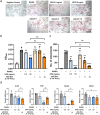
Inhibitory effect of ASC‐EVs on osteoclast differentiation of RAW264.7 cells. (a) TRAP staining of RAW264.7 cells after 6 days of incubation with RANKL containing medium supplemented with OPG (0.5 or 50 ng/ml) or ASC‐EVs (1 × 108 – 10 × 108 particles/ml) for 6 days. Scale bar, 200 μm. (b) TRAP activity in culture supernatants was assessed by measuring the optical density at 540 nm. (c) The number of TRAP‐positive multinucleated cells was observed under a light microscope and counted. (d) The gene expression associated with osteoclastogenesis in RAW264.7 cells was determined by qPCR. The relative gene expression was normalized to a housekeeping gene [glyceraldehyde 3‐phosphate dehydrogenase (Gapdh)] and expressed as the fold change compared to RANKL‐treated RAW264.7 cells. Data are mean ± SD (n = 3). *p < 0.05; **p < 0.01; ***p < 0.001; ****p < 0.0001; #p < 0.05; ##p < 0.01
FIGURE 6
FIGURE 6
OPG‐dependent anti‐osteoclastogenesis effects of ASC‐EVs. (a) Strategies to identify that anti‐osteoclastogenesis effects of ASC‐EVs are dependent on OPG. (b) OPG mRNA (TNFRSF11B) level in lentiviral control shRNA‐transduced ASCs (shCtrl ASCs) and lentiviral OPG shRNA‐transduced ASCs (shOPG ASCs). The relative OPG mRNA level was normalized to a housekeeping gene (18S) and expressed as the fold change compared to shCtrl ASCs. (c) Western blot representative images of shCtrl ASCs, shOPG ASCs, ASC‐EVs (Con‐EVs), and OPG‐depleted ASC‐EVs (OPG KD‐EVs). (d) The relative OPG protein expression was quantified in ASCs and ASC‐EVs using ImageJ software. The relative protein expression was normalized to Actin in ASCs and CD63 in ASC‐EVs. (e and f) TRAP staining representative images (e) and quantification of osteoclastogenesis in RAW264.7 cells (f). Scale bar, 500 μm. (g) The gene expression in RAW264.7 cells was determined by qPCR. The relative gene expression was normalized to a housekeeping gene (Gapdh) and expressed as the fold change compared to RANKL‐untreated RAW264.7 cells. Data are mean ± SD. ***p < 0.001; ****p < 0.0001; ####p < 0.0001
FIGURE 7
FIGURE 7
miRNA associated with osteoporosis present in ASC‐EVs. (a) Heatmap representing the mean RPM across replicates (n = 3). miRNAs in ASC‐EVs were quantified, and miRNA annotated reads were normalized to RPM. (b) The expression levels of miR‐21‐5p in RAW264.7 cells were determined by qPCR. The relative gene expression was normalized to miR‐16‐5p and Gapdh and expressed as the fold change compared to RANKL‐untreated RAW264.7 cells. (c) The miR‐21‐5p level in ASC‐EVs was measured by ddPCR and presented as copies/μL PCR. (d) The predicted target gene of miR‐21‐5p was determined by qPCR in RAW264.7 cells. The relative gene expression was normalized to a housekeeping gene (Gapdh) and expressed as the fold change compared to RANKL‐untreated RAW264.7 cells. (e) The expression levels of Acvr2a in RAW264.7 cells were determined by qPCR. The relative gene expression was normalized to a housekeeping gene (Gapdh) and expressed as the fold change compared to RANKL‐untreated RAW264.7 cells. (f) Predicted target sites of Acvr2a 3′UTR. Red letters showed the predicted pairing of the target region and miRNA. (g) The gene expression in RAW264.7 cells was determined by qPCR. The relative gene expression was normalized to a housekeeping gene (Gapdh) and expressed as the fold change compared to RANKL‐untreated RAW264.7 cells. (h) Cellular uptake of ASC‐EVs into RAW264.7 cells. Images of PKH‐67‐labeled ASC‐EVs (green) with DAPI (blue) were visualized by merging the confocal images. Scale bar, 20 μm. Data are mean ± SD. **p < 0.01; ***p < 0.001; ****p < 0.0001; #p < 0.05; ##p < 0.01
FIGURE 8
FIGURE 8
In vivo distribution of ADSC‐EVs in OVX mice. (a and b) Ex vivo bone distribution images (a) and quantification of fluorescence intensity (b) in OVX mice injected with Cy5.5 EVs or Cy5.5. Ex vivo images of bone tissues were harvested at 4 and 7 h after injection. (c–e) Ex vivo distribution images of major organs (heart, liver, spleen, kidney, and lung) (c) and quantification of fluorescence intensity at 4 h (d) and 7 h (e) after injection with Cy5.5 EVs or Cy5.5. Data are mean ± SD (n = 4). *p < 0.05; **p < 0.01; ***p < 0.001

References

    1. Antebi, B., Pelled, G., & Gazit, D. (2014). Stem cell therapy for osteoporosis. Current Osteoporosis Reports, 12(1), 41–47. 10.1007/s11914-013-0184-x - DOI - PubMed
    1. Biancone, L., Bruno, S., Deregibus, M. C., Tetta, C., & Camussi, G. (2012). Therapeutic potential of mesenchymal stem cell‐derived microvesicles. Nephrology, Dialysis, Transplantation, 27(8), 3037–3042. 10.1093/ndt/gfs168 - DOI - PubMed
    1. Bottani, M., Banfi, G., & Lombardi, G. (2019). Perspectives on MiRNAs as epigenetic markers in osteoporosis and bone fracture risk: A step forward in personalized diagnosis. Frontiers in Genetics, 10, 1044. 10.3389/fgene.2019.01044 - DOI - PMC - PubMed
    1. Boyce, B. F., & Xing, L. (2008). Functions of RANKL/RANK/OPG in bone modeling and remodeling. Archives of Biochemistry and Biophysics, 473(2), 139–146. 10.1016/j.abb.2008.03.018 - DOI - PMC - PubMed
    1. Bruno, S., Deregibus, M. C., & Camussi, G. (2015). The secretome of mesenchymal stromal cells: Role of extracellular vesicles in immunomodulation. Immunology Letters, 168(2), 154–158. 10.1016/j.imlet.2015.06.007 - DOI - PubMed

Publication types

Substances