. 2021 Oct 5;118(40):e2107535118.
doi: 10.1073/pnas.2107535118.

Nanoconfinement of microvilli alters gene expression and boosts T cell activation

Affiliations

Nanoconfinement of microvilli alters gene expression and boosts T cell activation

Morteza Aramesh et al. Proc Natl Acad Sci U S A. .

Erratum in

Abstract

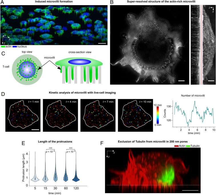
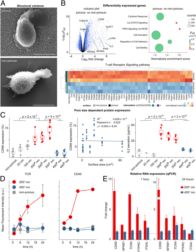
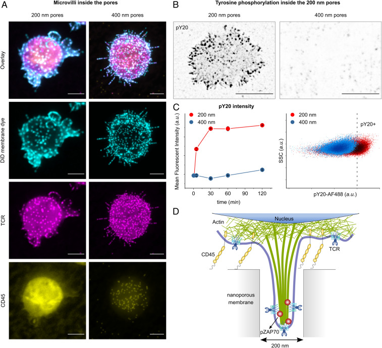
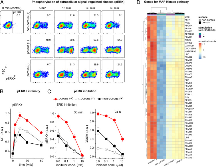
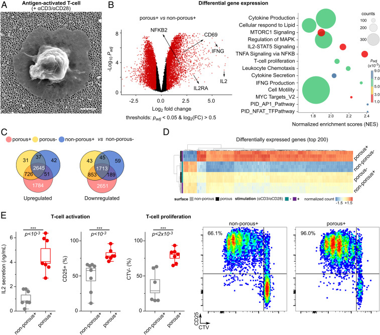
T cells sense and respond to their local environment at the nanoscale by forming small actin-rich protrusions, called microvilli, which play critical roles in signaling and antigen recognition, particularly at the interface with the antigen presenting cells. However, the mechanism by which microvilli contribute to cell signaling and activation is largely unknown. Here, we present a tunable engineered system that promotes microvilli formation and T cell signaling via physical stimuli. We discovered that nanoporous surfaces favored microvilli formation and markedly altered gene expression in T cells and promoted their activation. Mechanistically, confinement of microvilli inside of nanopores leads to size-dependent sorting of membrane-anchored proteins, specifically segregating CD45 phosphatases and T cell receptors (TCR) from the tip of the protrusions when microvilli are confined in 200-nm pores but not in 400-nm pores. Consequently, formation of TCR nanoclustered hotspots within 200-nm pores allows sustained and augmented signaling that prompts T cell activation even in the absence of TCR agonists. The synergistic combination of mechanical and biochemical signals on porous surfaces presents a straightforward strategy to investigate the role of microvilli in T cell signaling as well as to boost T cell activation and expansion for application in the growing field of adoptive immunotherapy.

Keywords: T cell microvilli; cell-surface interactions; immunoengineering; mechanobiology; nanoconfinement.


Conflict of interest statement

The authors declare no competing interest.

Figures

Fig. 1.
Fig. 1.
Microvilli protrusions in T cells on porous surfaces. (A) The confocal fluorescence microscopy images of Jurkat T cells on a porous AAO with 200-nm pore size (three-dimensional isometric projection). (Scale bar, 10 μm.) (B) A superresolution image of the actin cytoskeleton in a T cell using structured illumination microscopy. The protrusions are seen as a dotted structure in the top-view intensity projection and mostly concentrated around the cells’ periphery. (Scale bar, 1 μm.) (C) A schematic presentation of the actin-rich protrusions in T cells (not to scale). (D, Left) Snapshots of the actin protrusions in a live cell, obtained by fluorescent microscopy. The color corresponds to the lifetime of each protrusion. (Scale bar, 5 μm.) (Right) The number of protrusions in each frame, for the duration of 10 min. (E) The protrusions’ lengths were measured on fixed cells at the indicated time points. Violin plots show the distribution of the measured lengths (n = 20 cells). The P values were determined by two-sided Mann–Whitney U tests using R. (F) A cross-section view of a Jurkat T cell on a porous AAO with 200-nm pore size, captured by Airyscan confocal microscopy. The cells were stained with phalloidin (actin cytoskeleton) and antitubulin (microtubules), indicating that the microtubules were mostly excluded from entering the 200-nm pores. (Scale bar, 5 μm.)
Fig. 2.
Fig. 2.
Altered T cell gene expression in 200-nm porous substrates. (A) Scanning electron microscope images of nonstimulated primary human T cell on top of the porous (−) and nonporous (−) surfaces (without αCD3/CD28 coatings). (Scale bar, 4 μm.) (B) Nanoconfinement-induced alteration in the gene program of the primary human T cells after 4 h of seeding on the surfaces. Volcano plot shows differences in RNA expression of nonstimulated T cells on porous (−) versus nonporous (−) surfaces. Blue color indicates significance with adjusted P value <0.05 and log2 (fold changes) >0.5. Dot plot of gene set enrichment analysis shows differences in differentially enriched pathways of nonstimulated T cells on porous (−) versus nonporous (−) surfaces. The size of the circle corresponds to the gene counts (from the reference pathway), the color corresponds to the adjusted P value. (Bottom) Heatmap of the top 50 significantly up-regulated genes (adjusted P values <0.05) in the TCR signaling pathway, where genes in porous (−) cells were compared to nonporous (−) cells (n = 3 replicates, human primary T cells). The complete set of the significantly differentially expressed genes in TCR signaling pathway can be found in SI Appendix. (C) Jurkat T cells were seeded on porous and nonporous surfaces (without activating antibodies αCD3/CD28) with the indicated pore size (Table 1). CD69 and IL-2 were measured 24 h after activation by flow cytometry and ELISA, respectively. Box diagrams show data pooled from two or three independent experiments performed in triplicates. The scatter plot represents the pulled data from all experiments, plotted as surface area versus CD69 expression. The dashed line is the best linear fit to the data, which shows correlation between CD69 expression and increased surface area (Pearson’s r = −0.022). (D) The scatter plot shows the kinetics of the mean fluorescent intensity of TCR and CD45 of primary human T cells cultured on the indicated surfaces at different time points, measured by flow cytometry. Data represent one of two independent experiments performed in triplicates. Error bars are mean ± SD. (E) Relative RNA expression of selected genes measured by RT-qPCR. Primary human T cells were cultured on the indicated surfaces for 1 and 24 h. Fold expression is measured against the control sample (nonporous) and normalized by the reference gene RNA18S. The dashed line indicates fold expression =1, the expression of the genes in the control sample. The P values were determined by two-sided Mann–Whitney U tests in R.
Fig. 3.
Fig. 3.
Membrane-bound protein sorting in confined spaces. (A) The projection of confocal fluorescence microscopy images of the cell membrane (stained with DiD), TCR, and CD45, from fixed T cells on porous surfaces with (Left) 200-nm and (Right) 400-nm pore size, 30 min after seeding. (Scale bar, 5 μm.) (B) The projection of confocal fluorescence microscopy images of pY20, from fixed T cells on 200- and 400-nm pores, fixed 5 min after seeding. The fluorescent signal from inside of the pores is indicated by black dots. (Scale bar, 5 μm.) (C, Left) The scatter plot shows the kinetics of the mean fluorescent intensity of pY20 of primary human T cells cultured on the indicated surfaces at different time points, measured by flow cytometry. Data represent one of two independent experiments performed in triplicates. Error bars are mean ± SD. (Right) Representative dot plots of pY20 measured by flow cytometry on primary human T cells seeded on 200-nm (red) and 400-nm (blue) porous substrates, 30 min after seeding. (D) Schematics of the spatiotemporal segregation model induced by nanoconfinement in 200-nm pores.
Fig. 4.
Fig. 4.
ERK phosphorylation is augmented and sustained on the porous membranes. (A) Representative dot plots of phosphorylated ERK measured by flow cytometry on Jurkat T cells under the indicated conditions. Data are representative from two independent experiments performed in duplicates or triplicates. (B) The bar diagram shows the kinetics of the mean fluorescent intensity (MFI) of ERK-phosphorylated cells (pERK+) with the indicated treatment. Data represent one of two independent experiments performed in duplicates or triplicates. Error bars are mean ± SD. (C) Jurkat T cells were incubated with ERK inhibitor U0126 in different concentrations for 30 min before being activated on the indicated surfaces. Bar diagrams show percentages of pERK+ cells after 30 min and CD69+ cells after 24 h measured by flow cytometry (n = 3 replicates). Error bars are mean ± SD. (D) Heatmap of the top 50 significantly up-regulated genes in the MAP Kinase pathway of primary human T cells activated on the indicated surfaces for 4 h, adjusted P values <0.05. Genes were selected based on the comparison between porous (+) and nonporous (−) (n = 3 replicates). The complete set of the significantly differentially expressed genes in MAP Kinase pathway can be found in SI Appendix.
Fig. 5.
Fig. 5.
Boosted T cell activation and proliferation by combining nanotopographical and biochemical cues. (A) Scanning electron microscope image of an activated primary human T cell on top of the nanoporous membrane with 200-nm pore size. (Scale bar, 1 μm.) (B, Left) Volcano plot showing differences in RNA expression of T cells activated with antibodies on porous (+) versus nonporous (+) surfaces. Red color indicates significance with adjusted P value <0.05, and log2 (fold changes) >0.5. (Right) Pathway enrichment of differentially expressed genes in porous (+) cells versus nonporous (+) T cells shows increased stimulation of porous (+) T cells. The size of the circle corresponds to the gene counts (from the reference pathway), the color corresponds to the adjusted P value. (C) Venn diagrams of the differentially expressed genes (up-regulated and down-regulated) in three conditions [porous (+), porous (−), and nonporous (+) compared to the nonporous (−)]. (D) The top 200 significantly up-regulated genes in porous (+) compared to the nonporous (−), based on adjusted P values <0.05 (n = 3 replicates). +/− signs indicate the presence of activation antibodies (αCD3/CD28) on the surface. The complete set of the significantly differentially expressed genes can be found in SI Appendix. (E) Box diagrams show activation of human primary T cells on porous surfaces. IL-2 secretion was measured after 24 h, and CD25 expression was measured after 4 d. Three independent experiments were performed in duplicates or triplicates. The P values were determined by two-sided Mann–Whitney U tests in R. The proliferation assay (CTV, CellTrace Violet) was used to assess the expansion of the cells after 4 d. The graphs represent examples of the measurements by flow cytometry.

References

    1. Sage P. T., et al. ., Antigen recognition is facilitated by invadosome-like protrusions formed by memory/effector T cells. J. Immunol. 188, 3686–3699 (2012). - PMC - PubMed
    1. Ueda H., Morphew M. K., McIntosh J. R., Davis M. M., CD4+ T-cell synapses involve multiple distinct stages. Proc. Natl. Acad. Sci. U.S.A. 108, 17099–17104 (2011). - PMC - PubMed
    1. Leithner A., et al. ., Dendritic cell actin dynamics controls T cell priming efficiency at the immunological synapse. J Cell Biol 220, e202006081 (2021). - PMC - PubMed
    1. Aramesh M., et al. ., Functionalized bead assay to measure three-dimensional traction forces during T-cell activation. Nano Lett. 21, 507–514 (2021). - PubMed
    1. Jung Y., Wen L., Altman A., Ley K., CD45 pre-exclusion from the tips of microvilli establishes a phosphatase-free zone for early TCR triggering. Nat. Commun 12, 3872 (2021). - PMC - PubMed

Publication types

MeSH terms