. 2021 Nov;125(11):1523-1532.
doi: 10.1038/s41416-021-01561-0. Epub 2021 Oct 1.

APR-246 induces apoptosis and enhances chemo-sensitivity via activation of ROS and TAp73-Noxa signal in oesophageal squamous cell cancer with TP53 missense mutation

Affiliations

APR-246 induces apoptosis and enhances chemo-sensitivity via activation of ROS and TAp73-Noxa signal in oesophageal squamous cell cancer with TP53 missense mutation

Teruyuki Kobayashi et al. Br J Cancer. 2021 Nov.

Abstract

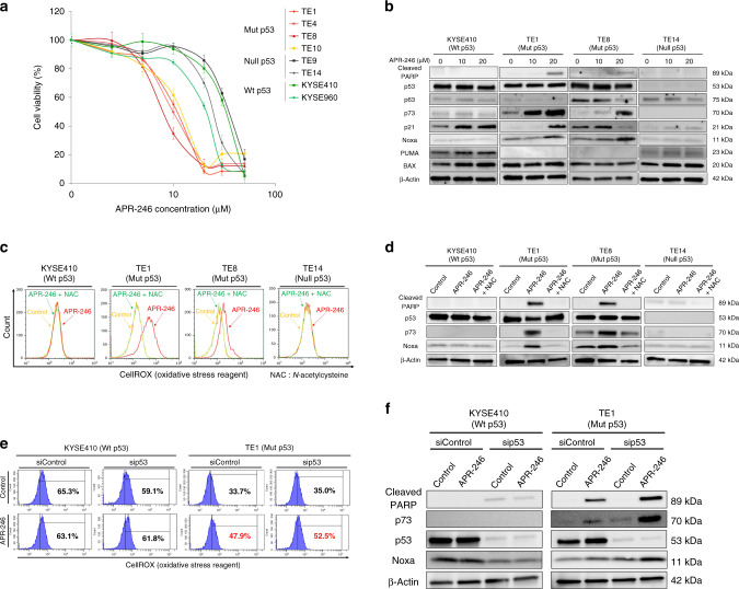
Background: Mutations in p53, identified in 90% of oesophageal squamous cell carcinoma (ESCC), are associated with unfavourable prognosis and chemo-resistance. APR-246 induces apoptosis by restoring transcriptional ability of mutant p53, and may be a promising therapeutic agent to overcome chemo-resistance in ESCC.

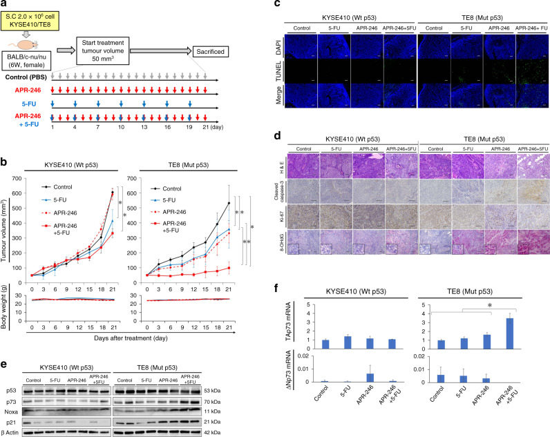
Methods: In ESCC cell lines differing in p53 status, we performed in vitro cell viability and apoptosis assays, evaluated reactive oxygen species (ROS) generation, and assessed signal changes by western blot after APR-246 administration with/without chemo-agent. Antitumour effects and signal changes were evaluated in in vivo experiments using xenograft and patient-derived xenograft (PDX) mouse models.

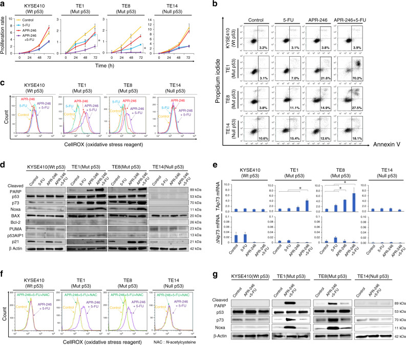
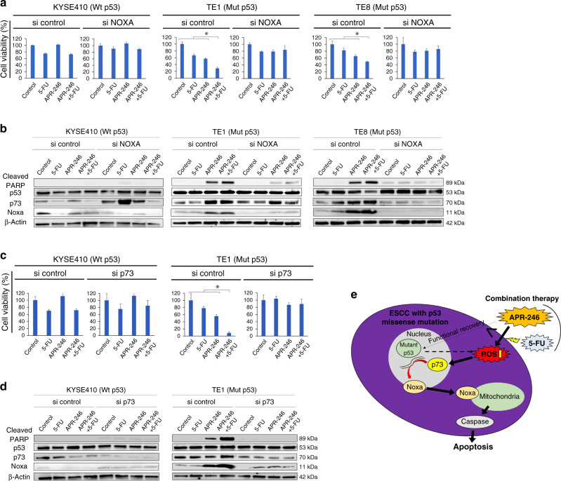
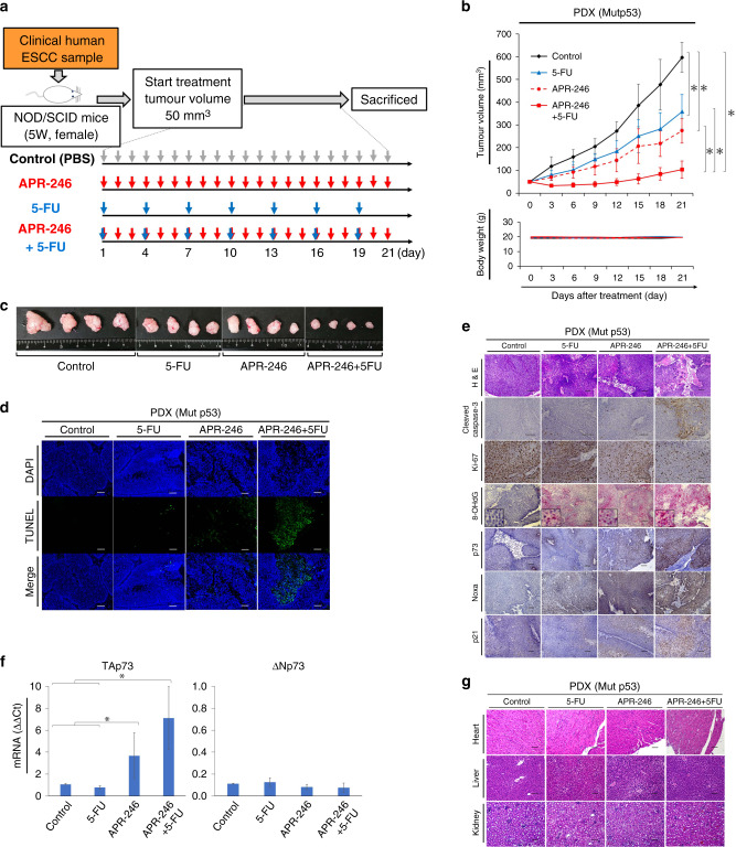
Results: APR-246 administration induced significant apoptosis by upregulating p73 and Noxa via ROS induction in ESCC cell lines harbouring p53 missense mutations. Moreover, APR-246 plus chemotherapy exerted combined antitumour effects in ESCC with p53 missense mutations. This effect was also mediated through enhanced ROS activity, leading to massive apoptosis via upregulation of p73 and Noxa. These findings were confirmed by xenograft and PDX models with p53 mutant ESCC.

Conclusion: APR-246 strongly induced apoptosis by inducing ROS activity and p73-Noxa signalling, specifically in ESCC with p53 missense mutation. This antitumour effect was further enhanced by combination with 5-FU, which we first confirmed in ESCC preclinical model.


Conflict of interest statement

The authors declare no competing interests.

Figures

Fig. 1
Fig. 1. APR-246 alone induces massive apoptosis via ROS induction leading to upregulation of p73-Noxa in ESCC with p53 missense mutation.
a APR-246 dose–response curve of ESCC cell lines after 48-h treatment with APR-246 as assessed by MTT cell viability assay. Error bars indicate standard deviation. b Western blot to assess relevant protein levels (cleaved PARP, p53, p63, p73, p21, Noxa, PUMA, and BAX) following treatment with APR-246 alone in ESCC cell lines (KYSE410, TE1, TE8, and TE14). c Flow cytometric data comparing ROS activities following administration of control, APR-246 alone, or APR-246 and N-acetylcysteine in ESCC cell lines (KYSE410, TE1, TE8, and TE14). d Comparison of relevant protein levels (cleaved PARP, p53, p73, and Noxa) under the same conditions as in c. e Comparison of ROS activities following administration of control or APR-246 alone in ESCC cell lines (KYSE410 and TE1) transfected with negative control or p53 siRNA. f Comparison of protein expressions under the same conditions as in e. P < 0.05. Wt wild type, Mut missense mutation, Null frameshift or nonsense mutation.
Fig. 2
Fig. 2. APR-246-induced apoptosis was further enhanced by its combination with 5-FU in ESCC with p53 missense mutation.
a Proliferation assay. ESCC cell lines (KYSE410, TE1, TE8, and TE14) were treated with PBS, APR-246 (10 μmol/L), 5-FU (10 μmol/L), or the combination of APR-246 (10 μmol/L) + 5-FU (10 μmol/L) for 72 h. b Apoptosis assay. Percentage of cells with positive annexin V staining after treatment with PBS, APR-246 (20 μmol/L), 5-FU (20 μmol/L), or the combination of APR-246 (20 μmol/L) + 5-FU (20 μmol/L) in ESCC cell lines (KYSE410, TE1, TE8, and TE14). c ROS activities under the same conditions as in b. d Western blotting to assess relevant protein levels (cleaved PARP, p53, p73, Noxa, BAX, Bcl-2, PUMA, p53AIP1, p21, and E2F-1) under the same condition as in b. e PCR results assessing TAp73 and ΔNp73 mRNA levels under the same condition as in e (ΔΔCt method). f Comparison of ROS activities after treatment with control, combination treatment (APR-246 20 μmol/L + 5-FU 20 μmol/L), or combination treatment plus N-acetylcysteine as assessed by flow cytometry in ESCC cell lines (KYSE410, TE1, TE8, and TE14). g Western blotting to assess relevant protein levels under the same conditions as in d. *P < 0.05. Wt wild type, Mut missense mutation, Null frameshift or nonsense mutation.
Fig. 3
Fig. 3. p73-Noxa upregulation as an essential pathway in combination therapy with APR-246 and 5-FU in ESCC with p53 missense mutation.
a Cell viability (%) after 48-h treatment with PBS, 5-FU (20 μmol/L), APR-246 (20 μmol/L), and the combination of APR-246 (20 μmol/L) + 5-FU (20 μmol/L) in ESCC cell lines (KYSE410, TE1, and TE8) transfected with si control or si NOXA. b Western blotting to assess relevant protein levels (cleaved PARP, p53, p73, and Noxa) under the same conditions as in e. c Cell viability (%) after 48-h treatment with PBS, 5-FU (20 μmol/L), APR-246 (20 μmol/L), and the combination of APR-246 (20 μmol/L) + 5-FU (20 μmol/L) in ESCC cell lines (KYSE410 and TE1) transfected with si control or si p73. d Western blotting to assess relevant protein levels (cleaved PARP, p53, p73, and Noxa) under the same condition as in c. e Schematic of the underlying mechanism through which APR-26, alone or in combination with a chemo-agent, induces significant apoptosis in ESCC with p53 missense mutation. *P < 0.05. Wt wild type, Mut missense mutation.
Fig. 4
Fig. 4. Combination treatment with APR-246 and 5-FU yields synergic anticancer effect in a xenograft mouse model of ESCC with p53 missense mutation.
a Schematic of the xenograft mouse model using ESCC cell lines with differing p53 status (KYSE410 versus TE8) treated for 21 days with PBS (control), 5-FU monotherapy (5 mg/kg every 3 days), APR-246 monotherapy (25 mg/kg daily), or the combination of 5-FU (5 mg/kg every 3 days) plus APR-246 (25 mg/kg daily). Treatment by intraperitoneal administration was started once tumour volume reached 50 mm3. Each group was sacrificed 21 days after the start of treatment. Each signal was evaluated using samples obtained 7 days after the treatment. b Tumour growth curve and weight change in each treatment group (n = 4). Tumour volume and body weight were measured every 3 days from the start of treatment. c Apoptosis associated with therapeutic effects in the tumour tissue of each treatment group as assessed by the TUNEL assay. d Comparison of immunostaining regarding apoptosis (cleaved caspase-3), cell proliferation ability (ki-67), and ROS activity between each treatment group. 8-OHdG staining: alkaline phosphatase-based immunostaining for the detection of ROS activity. Red staining indicates ROS activity, while purple staining shows haematoxylin and eosin. e Western blotting to assess relevant protein levels (p53, p73, Noxa, and p21) in tumour tissues of each treatment group. f PCR assay to measure the mRNA expression levels of TAp73 and ΔNp73 (PCR ΔΔCt method) in tumour tissues of each treatment group. Scale bars, 100 μm. *P < 0.05. Wt wild type, Mut missense mutation.
Fig. 5
Fig. 5. PDX mouse model confirmed ROS-TAp73-Noxa-mediated apoptosis by treatment with APR-246 plus 5-FU without any adverse events.
a Schematic of the PDX mouse model using tumour tissue that was confirmed to include p53 missense mutation [p.Val118Phe (exon5) and p.Gly206Val (exon7)] by Sanger sequencing, from a resected specimen from an ESCC patient. Mice were treated with PBS (control), 5-FU monotherapy (5 mg/kg every 3 days), APR-246 monotherapy (25 mg/kg daily), or combined therapy for 21 days. Treatment by intraperitoneal administration was started once tumour volume reached 50 mm3. Each group was sacrificed 21 days after the start of treatment. Samples were obtained on Day 7 for signal evaluation and on Day 21 to evaluate treatment effects on each organ. b Tumour growth curve and weight change in each treatment group (n = 4). Tumour volume and body weight were measured every 3 days from the start of treatment. c Macroscopic findings of resected tumours in each treatment group. d Apoptosis associated with therapeutic effects in tumour tissue from the PDX mouse model of each group, as assessed by TUNEL assay. e Comparison of immunostaining regarding apoptosis (cleaved caspase-3), cell proliferation ability (ki-67), ROS activity (8OH-dG), p73, Noxa, and p21 in tumour tissues of each treatment group. f PCR assay of mRNA expression of TAp73 and ΔNp73 (PCR ΔΔCt method) in tumour tissues of each treatment group. g Haematoxylin and eosin staining of heart, kidney, and liver tissue from each treatment group. Scale bars, 100 μm. *P < 0.05. Mut missense mutation.

References

    1. Colvin H, Mizushima T, Eguchi H, Takiguchi S, Doki Y, Mori M. Gastroenterological surgery in Japan: the past, the present and the future. Ann Gastroenterol Surg. 2017;1:5–10. - PMC - PubMed
    1. Ando N, Kato H, Igaki H, Shinoda M, Ozawa S, Shimizu H, et al. A randomized trial comparing postoperative adjuvant chemotherapy with cisplatin and 5-fluorouracil versus preoperative chemotherapy for localized advanced squamous cell carcinoma of the thoracic esophagus (JCOG9907) Ann Surg Oncol. 2012;19:68–74. - PubMed
    1. Beroud C, Soussi T. The UMD-p53 database: new mutations and analysis tools. Hum Mutat. 2003;21:176–81. - PubMed
    1. Sawada G, Niida A, Uchi R, Hirata H, Shimamura T, Suzuki Y, et al. Genomic landscape of esophageal squamous cell carcinoma in a Japanese population. Gastroenterology. 2016;150:1171–82. - PubMed
    1. Yamasaki M, Miyata H, Fujiwara Y, Takiguchi S, Nakajima K, Nishida T, et al. p53 genotype predicts response to chemotherapy in patients with squamous cell carcinoma of the esophagus. Ann Surg Oncol. 2010;17:634–42. - PubMed

MeSH terms