

Brain aging mechanisms with mechanical manifestations
- PMID: 34600936
- PMCID: PMC8627478
- DOI: 10.1016/j.mad.2021.111575
Brain aging mechanisms with mechanical manifestations
Abstract
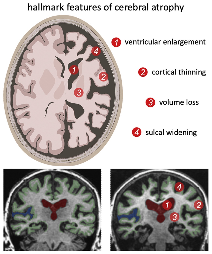
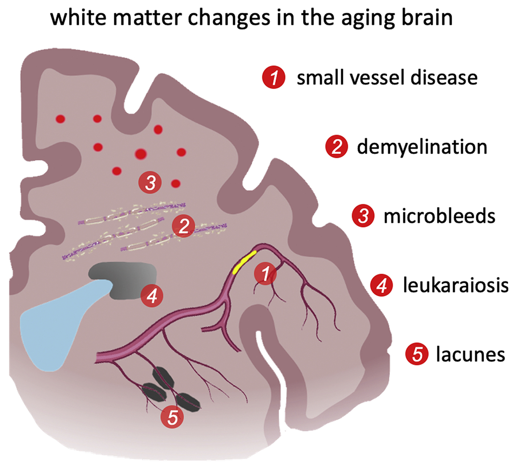
Brain aging is a complex process that affects everything from the subcellular to the organ level, begins early in life, and accelerates with age. Morphologically, brain aging is primarily characterized by brain volume loss, cortical thinning, white matter degradation, loss of gyrification, and ventricular enlargement. Pathophysiologically, brain aging is associated with neuron cell shrinking, dendritic degeneration, demyelination, small vessel disease, metabolic slowing, microglial activation, and the formation of white matter lesions. In recent years, the mechanics community has demonstrated increasing interest in modeling the brain's (bio)mechanical behavior and uses constitutive modeling to predict shape changes of anatomically accurate finite element brain models in health and disease. Here, we pursue two objectives. First, we review existing imaging-based data on white and gray matter atrophy rates and organ-level aging patterns. This data is required to calibrate and validate constitutive brain models. Second, we review the most critical cell- and tissue-level aging mechanisms that drive white and gray matter changes. We focuse on aging mechanisms that ultimately manifest as organ-level shape changes based on the idea that the integration of imaging and mechanical modeling may help identify the tipping point when normal aging ends and pathological neurodegeneration begins.
Keywords: Brain aging mechanisms; Cerebral atrophy; Gray and white matter changes; Morphological changes; Vascular changes.
Copyright © 2021 The Author(s). Published by Elsevier B.V. All rights reserved.
Figures
References
-
- Abe O, Yamasue H, Aoki S, Suga M, Yamada H, Kasai K, Masutani Y, Kato N, Kato N, Ohtomo K, 2008. Aging in the cns: comparison of gray/white matter volume and diffusion tensor data. Neurobiol. Aging 29, 102–116. - PubMed
-
- Allen JS, Bruss J, Brown CK, Damasio H, 2005. Normal neuroanatomical variation due to age: the major lobes and a parcellation of the temporal region. Neurobiol. Aging 26, 1245–1260. - PubMed
-
- Ambarki K, Israelsson H, Wåhlin A, Birgander R, Eklund A, Malm J, 2010. Brain ventricular size in healthy elderly: comparison between evans index and volume measurement. Neurosurgery 67, 94–99. - PubMed
Publication types
MeSH terms
Grants and funding
LinkOut - more resources
Full Text Sources
Medical
