. 2022 Mar;16(3):788-800.
doi: 10.1038/s41396-021-01121-7. Epub 2021 Oct 2.

Molecular basis of a bacterial-amphibian symbiosis revealed by comparative genomics, modeling, and functional testing

Affiliations

Molecular basis of a bacterial-amphibian symbiosis revealed by comparative genomics, modeling, and functional testing

Andrés E Brunetti et al. ISME J. 2022 Mar.

Abstract

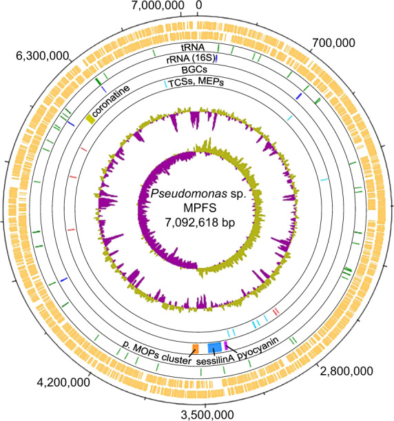
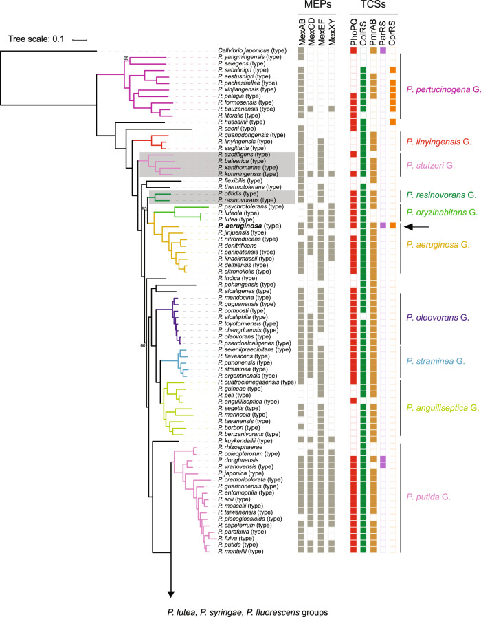
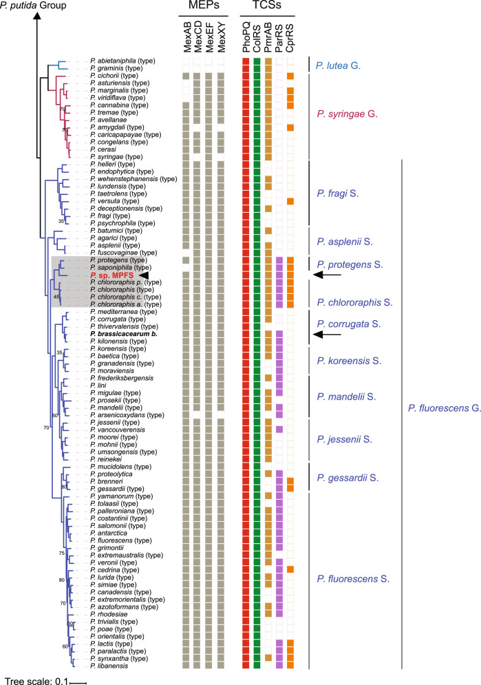
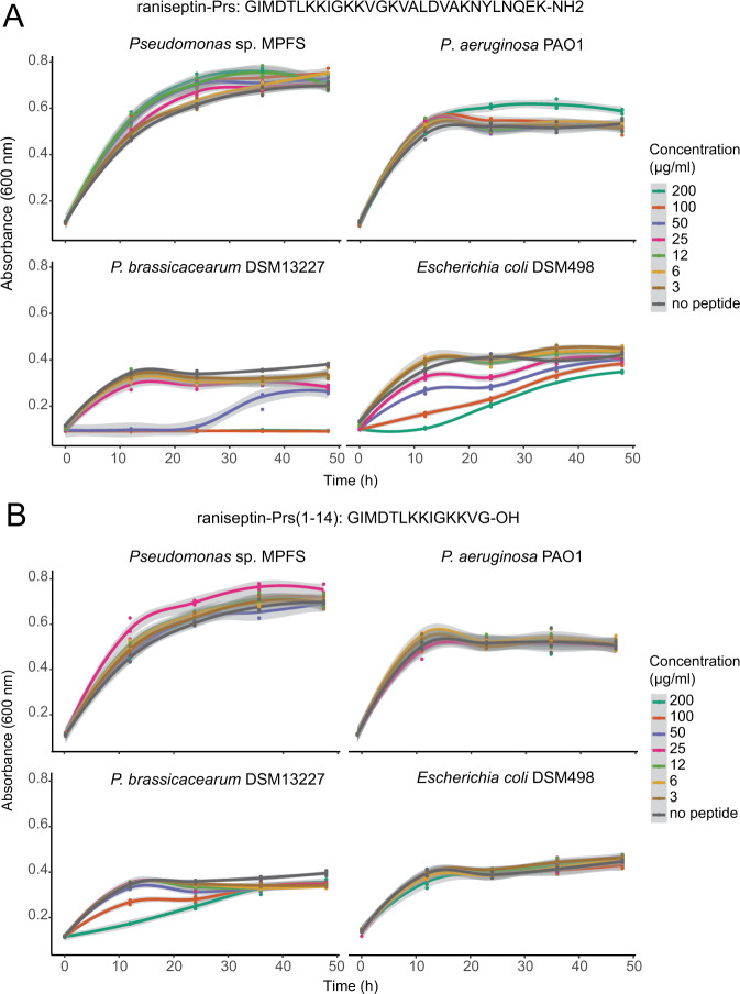
The molecular bases for the symbiosis of the amphibian skin microbiome with its host are poorly understood. Here, we used the odor-producer Pseudomonas sp. MPFS and the treefrog Boana prasina as a model to explore bacterial genome determinants and the resulting mechanisms facilitating symbiosis. Pseudomonas sp. MPFS and its closest relatives, within a new clade of the P. fluoresens Group, have large genomes and were isolated from fishes and plants, suggesting environmental plasticity. We annotated 16 biosynthetic gene clusters from the complete genome sequence of this strain, including those encoding the synthesis of compounds with known antifungal activity and of odorous methoxypyrazines that likely mediate sexual interactions in Boana prasina. Comparative genomics of Pseudomonas also revealed that Pseudomonas sp. MPFS and its closest relatives have acquired specific resistance mechanisms against host antimicrobial peptides (AMPs), specifically two extra copies of a multidrug efflux pump and the same two-component regulatory systems known to trigger adaptive resistance to AMPs in P. aeruginosa. Subsequent molecular modeling indicated that these regulatory systems interact with an AMP identified in Boana prasina through the highly acidic surfaces of the proteins comprising their sensory domains. In agreement with a symbiotic relationship and a highly selective antibacterial function, this AMP did not inhibit the growth of Pseudomonas sp. MPFS but inhibited the growth of another Pseudomonas species and Escherichia coli in laboratory tests. This study provides deeper insights into the molecular interaction of the bacteria-amphibian symbiosis and highlights the role of specific adaptive resistance toward AMPs of the hosts.


Conflict of interest statement

The authors declare no competing interests.

Figures

Fig. 1
Fig. 1. Circular plot of the 7.1 Mbp Pseudomonas sp. MPFS chromosome and COG functional categories.
The eight circles from the outermost to the innermost depict either the location of different genes or general genome features: circles 1 and 2 (orange) predicted coding sequences on the positive and negative strands, respectively; circles 3 (green) and 4 (blue), tRNA and rRNA genes, respectively; circle 5, biosynthetic clusters (BGCs) with high similarities (≥75%) to BGCs with known function, and a nonribosomal peptide synthetase putatively involved in the synthesis of methoxypyrazines (p. MOPs cluster, orange); circle 6, the five two-component system genes (TCSs, red) and eight multidrug efflux pumps (MEPs, cyan) known to mediate adaptive resistance against antimicrobial peptides (AMPs) in Pseudomonas aeruginosa, and; circle 7 and 8, %G+C and GC skew, respectively.
Fig. 2
Fig. 2. Phylogenetic relationships of Pseudomonas obtained with maximum likelihood based on protein sequence alignments of orthologous genes from 141 type strains (type), 17 non-type strains, and the yet undescribed Pseudomonas sp. MPFS from this study (arrowhead, red, bold).
Cellvibrio japonicus was included as an outgroup. Nodes without numbers indicate bootstrap values higher than 95%. Branch colors depict Pseudomonas spp. groups (G.) and P. fluorescens subgroups (S.), and portions of the tree highlighted in gray depict groups or subgroups that differ from previous genome-based phylogenies. Columns on the right indicate the presence/absence of genes encoding for multidrug efflux pumps (MEPs) and two-component sensory systems (TCSs) known to participate in adaptive resistance in P. aeruginosa. Arrows and names in bold font indicate the strains selected for inhibition experiments.
Fig. 3
Fig. 3. Phylogenomic tree showing the position of Pseudomonas sp. MPFS in the most closely related clades and overview of ecological niches used by different clades of Pseudomonas.
A Based on the tree inferred with FastME 2.1.4 using the TYGS platform and on the genome BLAST distance phylogeny method (GBDP), we defined P. piscis Subgroup (S.) as a new clade within the P. fluorescens Group. Branch lengths are scaled in terms of GBDP distance formula d5, and the numbers on branches are GBDP pseudo-bootstrap support values from 100 replications. Different square colors on the right panel depict whether the strains belong to a given species and subspecies cluster, whereas the color intensity of the G+C content depicts their relative values. B A simplified list depicting known biological sources from where different strains (str.) from the Pseudomonas groups and Pseudomonas fluorescens subgroups included in this study were identified. Red rectangles on symbols depict pathogenic interactions. GS genome size, fl free-living strains, sy str symbiotic strains.
Fig. 4
Fig. 4. Biosynthetic gene clusters (BGCs) from Pseudomonas sp. MPFS and from strains within the P. piscis, P. protegens, and P. chlororaphis subgroups with reliable secondary metabolite assignment (sequence similarity ≥75% with clusters of known function).
A Three BGC types, phenazine (top), nonribosomal peptide synthetase (NRPS) (second from the top), and NRPS-like (third from the top), have high sequence similarities to clusters occurring in other Pseudomonas strains (denoted on the right of the panel). In addition, a dimodular NRPS putatively involved in the synthesis of methoxypyrazines was identified (bottom). B Total number of BGCs and presence/absence of BGCs with reliable secondary metabolite assignment. Information on the presence/absence of antifungal activity for these compounds was obtained from the literature as detailed in Supplementary Table S8.
Fig. 5
Fig. 5. Bacterial growth curves of three Pseudomonas strains and Escherichia coli exposed to different concentrations of the cationic antimicrobial peptide raniseptin-Prs from Boana prasina.
A full length peptide, and B N-cleaved peptide at the Gly14-Lys15 position. Shaded areas in each curve represent 95% confidence intervals.
Fig. 6
Fig. 6. Tertiary structure model of the sensory periplasmatic domain of the sensory histidine kinases PhoQ and CprS from Pseudomonas aeruginosa strain PAO1 (left panel) and Pseudomonas sp. MPFS (right panel), and modeled docking with the peptide raniseptin-Prs.
A Both sensory domains form a PAS-like fold. B Net charge at pH = 7.0 and electrostatic potential (±2kT/e) visualized over molecular surface. The red to blue color gradient depicts the negative to positive potentials, respectively. The Asp and Glu residues near the expected membrane region are depicted for all sensory domains. C Structure of complexes of raniseptin-Prs with PhoQ and CprS and respective scores, in arbitrary energy units (a.e.u.), obtained by docking. The hydrophobic residues of raniseptin-Prs are shown as gray dots. Cytoplasmic membrane is represented only for reference.

References

    1. Gallo RL, Hooper LV. Epithelial antimicrobial defence of the skin and intestine. Nat Rev Immunol. 2012;12:503–16. - PMC - PubMed
    1. Erin Chen Y, Fischbach MA, Belkaid Y. Skin microbiota-host interactions. Nature. 2018;553:427–36. - PMC - PubMed
    1. Toledo RC, Jared C. Cutaneous granular glands and amphibian venoms. Comp Biochem Physiol A Physiol. 1995;111:1–29.
    1. Walke JB, Becker MH, Loftus SC, House LL, Cormier G, Jensen RV, et al. Amphibian skin may select for rare environmental microbes. ISME J. 2014;8:2207–17. - PMC - PubMed
    1. Jani AJ, Briggs CJ. Host and aquatic environment shape the amphibian skin microbiome but effects on downstream resistance to the pathogen Batrachochytrium dendrobatidis are variable. Front Microbiol. 2018;9:487. - PMC - PubMed

Publication types