

Abdominal imaging findings on computed tomography in patients acutely infected with SARS-CoV-2: what are the findings?
- PMID: 34601700
- PMCID: PMC8487449
- DOI: 10.1007/s10140-021-01986-3
Abdominal imaging findings on computed tomography in patients acutely infected with SARS-CoV-2: what are the findings?
Abstract
Objectives: To investigate what findings are new on contrast-enhanced abdominopelvic CT in patients infected with SARS-CoV-2.
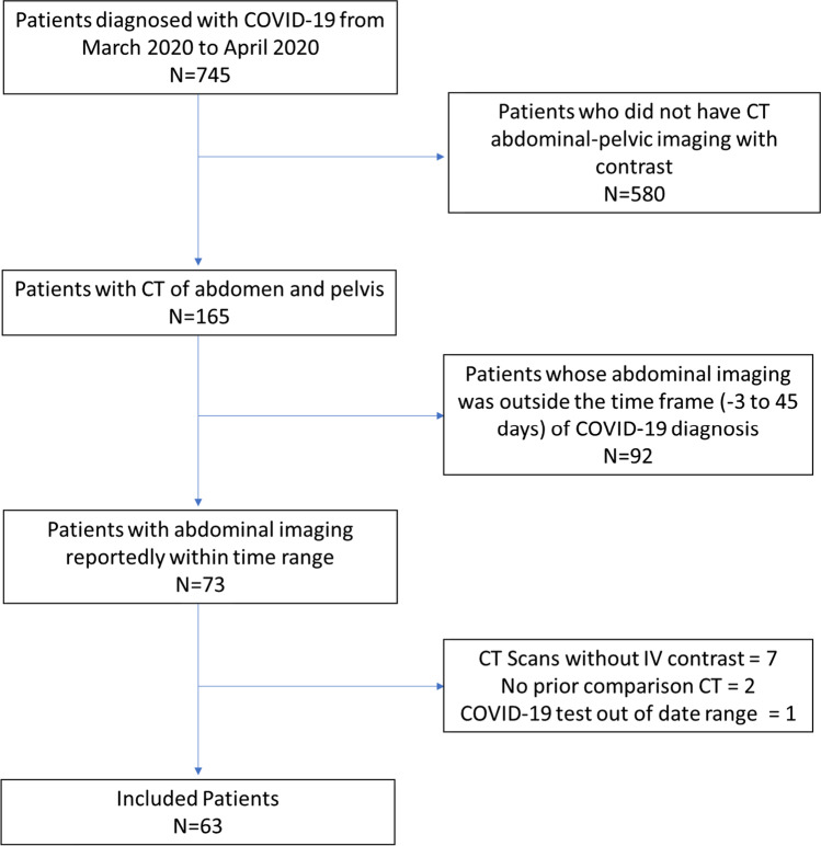
Methods: Contrast-enhanced CT of the abdomen and pelvis of patients with COVID-19 at a tertiary oncologic center acquired over a 2-month period were reviewed independently by two readers and scored for new imaging abnormalities compared with a prior scan. CT scans were included if the study was performed between - 3 and 45 days from the time of COVID-19 diagnosis. Clinical information was gathered from the medical records.
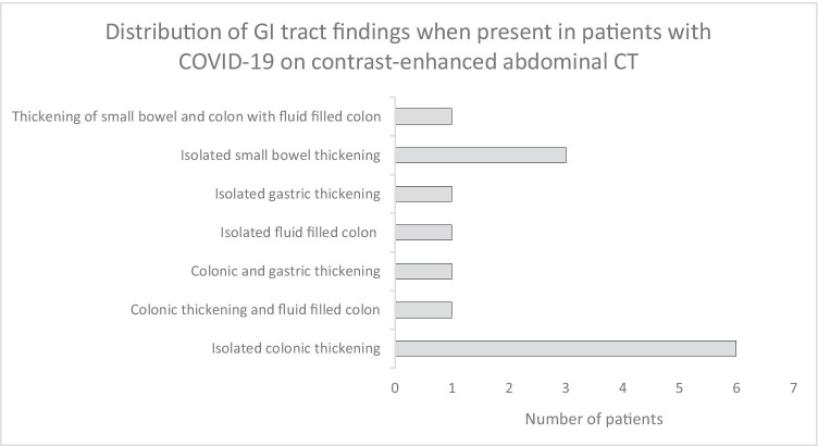
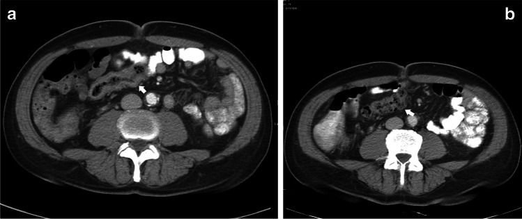
Results: A total of 63 patients (34 male, 29 female; mean age 60.6 years, range 24.4-85.0 years) were included in this retrospective cross-sectional study. Aside from new ground glass opacities seen at the lung bases (29/63, 46.0%), the most common findings were new thickening of the stomach, small bowel or colon or fluid-filled colon (14/63, 22.2%), new small volume ascites (7/63, 14.3%), gallbladder distention in those without prior cholecystectomy (3/43, 7.0%), and single cases each of acute pancreatitis (1/63, 1.6%) as well as new portal vein thrombosis (1/63, 1.6%).
Conclusion: Aside from lung base ground glass opacities, the most common new imaging abnormality on abdominopelvic CT in patients with COVID-19 finding in our cohort was abnormalities of the gastrointestinal tract, followed by small volume ascites, gallbladder distention, and isolated cases of pancreatitis and portal vein thrombosis. These findings overlap with those previously reported that did not have a prior scan for comparison, and provide supportive evidence that some of these findings may be related to SARS-CoV-2 infection.
Keywords: COVID-19; Computed tomography; Coronavirus; Enterocolitis; SARS-CoV-2.
© 2021. American Society of Emergency Radiology.
Conflict of interest statement
The authors declare that they have no conflict of interest.
Figures


References
-
- Organization WH (2021) Numbers at a glance. https://www.who.int/emergencies/diseases/novel-coronavirus-2019. Accessed 23 July 2021
-
- Bai C, Chotirmall SH, Rello J, Alba GA, Ginns LC, Krishnan JA, et al. Updated guidance on the management of COVID-19: from an American thoracic society/European respiratory society coordinated international task force (29 July 2020) Eur Respir Rev. 2020 doi: 10.1183/16000617.0287-2020. - DOI - PMC - PubMed
MeSH terms
Grants and funding
LinkOut - more resources
Full Text Sources
Medical
Miscellaneous
