

doi: 10.1590/0100-3984.2020.0137.
MV-Flow and LumiFlow: new Doppler tools for evaluating the microvasculature of the fetal head
Affiliations
- PMID: 34602672
- PMCID: PMC8475164
- DOI: 10.1590/0100-3984.2020.0137
Item in Clipboard
MV-Flow and LumiFlow: new Doppler tools for evaluating the microvasculature of the fetal head
Radiol Bras.
2021 Sep-Oct.
No abstract available
Figures
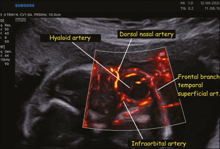
Coronal view of the fetal face, obtained with MV-Flow and LumiFlow at 28 weeks of gestation, showing the orbital arteries. art., artery.
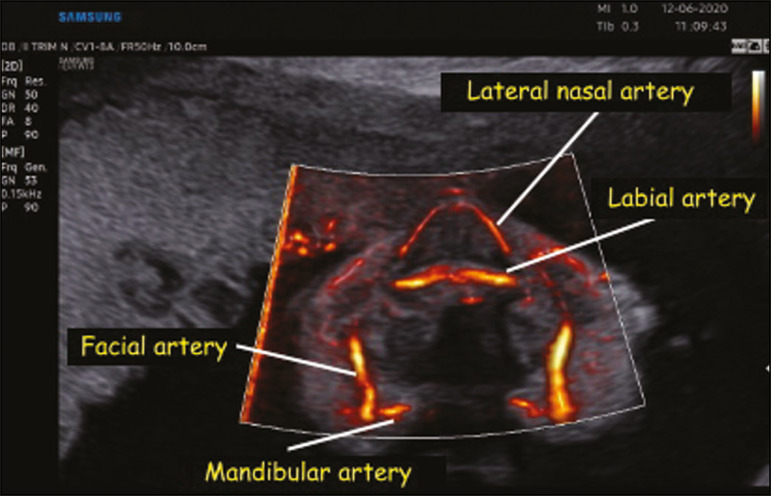
Coronal view of the fetal face at the level of the alveolar portion of the palate, obtained with MV-Flow and LumiFlow at 28 weeks of gestation, showing the facial, labial, nasal, and mandibular arteries.
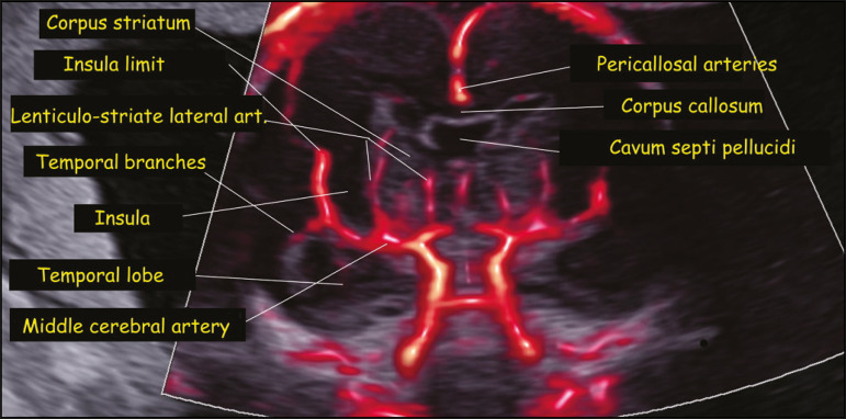
Coronal view of the fetal central nervous system at the level of the corpus callosum, obtained with MV-Flow and LumiFlow at 22 weeks of gestation, showing the cerebral microcirculation. art., artery.
Similar articles
-
MV-Flow and LumiFlow: new Doppler tools for the visualization of fetal blood vessels.Radiol Bras. 2021 Jul-Aug;54(4):277-278. doi: 10.1590/0100-3984.2020.0109. Radiol Bras. 2021. PMID: 34393297 Free PMC article. No abstract available.
-
MV-Flow and LumiFlow: a new Doppler tool for assessing the development of fetal brain vascularization in late-first/early-second trimester of pregnancy.J Ultrason. 2021 Aug 16;21(86):e258-e259. doi: 10.15557/JoU.2021.0042. Epub 2021 Sep 9. J Ultrason. 2021. PMID: 34540283 Free PMC article. No abstract available.
-
Evaluation of a new 3-dimensional color Doppler flow method to quantify flow across the mitral valve and in the left ventricular outflow tract: an in vitro study.J Ultrasound Med. 2014 Feb;33(2):265-71. doi: 10.7863/ultra.33.2.265. J Ultrasound Med. 2014. PMID: 24449729
-
[Contribution of Doppler exploration of ductus venosus flow].J Gynecol Obstet Biol Reprod (Paris). 2002 Feb;31(1 Suppl):2S64-9. J Gynecol Obstet Biol Reprod (Paris). 2002. PMID: 11973522 Review. French.
-
[Obstetric Doppler--justified i high-risk pregnancies].Lakartidningen. 1998 Sep 30;95(40):4360-4. Lakartidningen. 1998. PMID: 9800456 Review. Swedish.
Cited by
-
Application of O-RADS US combined with MV-Flow to diagnose ovarian-adnexal tumors.Ultrasonography. 2024 Jan;43(1):15-24. doi: 10.14366/usg.23061. Epub 2023 Aug 25. Ultrasonography. 2024. PMID: 38061878 Free PMC article.
-
Update on newer ultrasound systems to study the microvasculature.Radiol Med. 2025 Aug;130(8):1283-1296. doi: 10.1007/s11547-025-02035-6. Epub 2025 Jun 25. Radiol Med. 2025. PMID: 40560337 Review.
-
Alterations on magnetic resonance imaging of the neonatal brain: correlations with prenatal risk factors and transfontanellar ultrasound findings.Radiol Bras. 2022 Sep-Oct;55(5):280-285. doi: 10.1590/0100-3984.2021.0149-en. Radiol Bras. 2022. PMID: 36320373 Free PMC article.
-
Microvascular assessment of fascio-cutaneous flaps by ultrasound: A large animal study.Front Physiol. 2022 Dec 15;13:1063240. doi: 10.3389/fphys.2022.1063240. eCollection 2022. Front Physiol. 2022. PMID: 36589429 Free PMC article.
-
Effectiveness of microvascular flow imaging for radiofrequency ablation in recurrent thyroid cancer: comparison with power Doppler imaging.Eur Radiol. 2025 Feb;35(2):597-607. doi: 10.1007/s00330-024-10977-0. Epub 2024 Jul 23. Eur Radiol. 2025. PMID: 39042304
References
-
- Ohlmann P, Jung F, Mrowietz C, et al. Peripheral microcirculation during pregnancy and in women with pregnancy induced hypertension. Clin Hemorheol Microcirc. 2001;24:183–191. - PubMed
-
- Chang CH, Yu CH, Ko HC, et al. Three-dimensional power Doppler ultrasound for the assessment of the fetal brain blood flow in normal gestation. Ultrasound Med Biol. 2003;29:1273–1279. - PubMed
-
- Volpe P, Persico N, Fanelli T, et al. Prospective detection and differential diagnosis of cystic posterior fossa anomalies by assessing posterior brain at 11-14 weeks. Am J Obstet Gynecol MFM. 2019;1:173–181. - PubMed
-
- Dall'Asta A, Grisolia G, Volpe N, et al. Prenatal visualisation of the torcular herophili by means of a Doppler technology highly sensitive for low-velocity flow in the expert assessment of the posterior fossa: a prospective study. BJOG. 2021;128:347–352. - PubMed
-
- Konje JC, Abrams K, Bell SC, et al. The application of color power angiography to the longitudinal quantification of blood flow volume in the fetal middle cerebral arteries, ascending aorta, descending aorta, and renal arteries during gestation. Am J Obstet Gynecol. 2000;182:393–400. - PubMed
LinkOut - more resources
Full Text Sources