

doi: 10.1590/0100-3984.2020.0137.
MV-Flow and LumiFlow: new Doppler tools for evaluating the microvasculature of the fetal head
Affiliations
- PMID: 34602672
- PMCID: PMC8475164
- DOI: 10.1590/0100-3984.2020.0137
Item in Clipboard
MV-Flow and LumiFlow: new Doppler tools for evaluating the microvasculature of the fetal head
Radiol Bras.
2021 Sep-Oct.
No abstract available
Figures
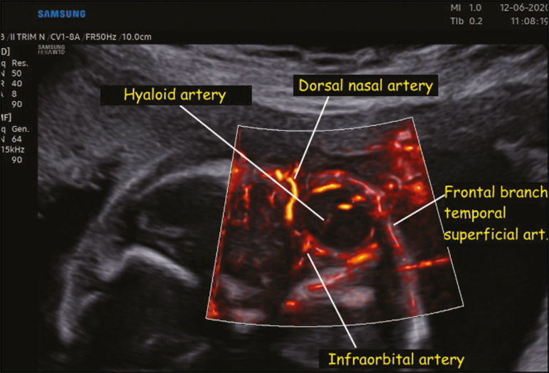
Coronal view of the fetal face, obtained with MV-Flow and LumiFlow at 28 weeks of gestation, showing the orbital arteries. art., artery.
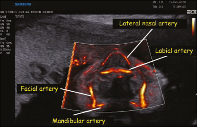
Coronal view of the fetal face at the level of the alveolar portion of the palate, obtained with MV-Flow and LumiFlow at 28 weeks of gestation, showing the facial, labial, nasal, and mandibular arteries.
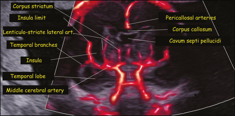
Coronal view of the fetal central nervous system at the level of the corpus callosum, obtained with MV-Flow and LumiFlow at 22 weeks of gestation, showing the cerebral microcirculation. art., artery.
References
-
- Ohlmann P, Jung F, Mrowietz C, et al. Peripheral microcirculation during pregnancy and in women with pregnancy induced hypertension. Clin Hemorheol Microcirc. 2001;24:183–191. - PubMed
-
- Chang CH, Yu CH, Ko HC, et al. Three-dimensional power Doppler ultrasound for the assessment of the fetal brain blood flow in normal gestation. Ultrasound Med Biol. 2003;29:1273–1279. - PubMed
-
- Volpe P, Persico N, Fanelli T, et al. Prospective detection and differential diagnosis of cystic posterior fossa anomalies by assessing posterior brain at 11-14 weeks. Am J Obstet Gynecol MFM. 2019;1:173–181. - PubMed
-
- Dall'Asta A, Grisolia G, Volpe N, et al. Prenatal visualisation of the torcular herophili by means of a Doppler technology highly sensitive for low-velocity flow in the expert assessment of the posterior fossa: a prospective study. BJOG. 2021;128:347–352. - PubMed
-
- Konje JC, Abrams K, Bell SC, et al. The application of color power angiography to the longitudinal quantification of blood flow volume in the fetal middle cerebral arteries, ascending aorta, descending aorta, and renal arteries during gestation. Am J Obstet Gynecol. 2000;182:393–400. - PubMed
LinkOut - more resources
Full Text Sources