Intravascular Papillary Endothelial Hyperplasia: Case Report of a Recurrent Masson's Tumor of the Finger and Review of Literature
- PMID: 34602798
- PMCID: PMC8463135
- DOI: 10.1055/s-0039-3401381
Intravascular Papillary Endothelial Hyperplasia: Case Report of a Recurrent Masson's Tumor of the Finger and Review of Literature
Abstract
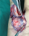
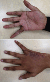
Intravascular papillary endothelial hyperplasia (IPEH), often referred to as Masson's Tumor, is an uncommon yet benign vascular disease of the skin and subcutaneous tissues. It usually arises within a blood vessel, but is considered to be a non-neoplastic reactive endothelial proliferation commonly associated with vascular injury. Although it is rare, knowledge of this disease is important as it may mimic other benign and malignant tumors, especially angiosarcoma, which may lead to unnecessary aggressive management. Typically, IPEHs are asymptomatic and are slow growing soft-tissue masses with extremely low-recurrence rates. In this article, we describe a 19-year-old male with a recurrence of Masson's Tumor over the right little finger within 2 months of a routine excision of the lesion. We also present accompanying multimodality clinical, radiological, and pathological imaging. This case illustrates the innocuous nature of the initial lesion easily mistaken for a hemangioma. Awareness of the possibility of a recurrence of a Masson's Tumor is important for clinicians to rule out the presence of malignant vascular lesions.
Keywords: Masson’s Tumor; finger; intravascular papillary endothelial hyperplasia; recurrence.
Society of Indian Hand & Microsurgeons. All rights reserved. Thieme Medical and Scientific Publishers Pvt. Ltd., A-12, 2nd Floor, Sector 2, Noida-201301 UP, India.
Conflict of interest statement
Conflict of Interest None declared.
Figures


References
-
- Masson P. Hemangioendotheliome vegetant intravasculaire. Bull Soc Anat Paris. 1923;93:517–523.
-
- Henschen F. L’endovasculite proliférante thrombopoiétique dans la lésion vasculaire locale. Ann Anat Pathol (Paris) 1932;9:113–121.
-
- Erol O, Ozçakar L, Uygur F, Keçik A, Ozkaya O. Intravascular papillary endothelial hyperplasia in the finger: not a premier diagnosis. J Cutan Pathol. 2007;34(10):806–807. - PubMed
-
- Clearkin K P, Enzinger F M. Intravascular papillary endothelial hyperplasia. Arch Pathol Lab Med. 1976;100(08):441–444. - PubMed
-
- Levere S M, Barsky S H, Meals R A. Intravascular papillary endothelial hyperplasia: a neoplastic “actor” representing an exaggerated attempt at recanalization mediated by basic fibroblast growth factor. J Hand Surg Am. 1994;19(04):559–564. - PubMed
Publication types
LinkOut - more resources
Full Text Sources

