. 2021 Sep 16:12:620058.
doi: 10.3389/fphys.2021.620058. eCollection 2021.

Fetal Sex and Fetal Environment Interact to Alter Diameter, Myogenic Tone, and Contractile Response to Thromboxane Analog in Rat Umbilical Cord Vessels

Affiliations

Fetal Sex and Fetal Environment Interact to Alter Diameter, Myogenic Tone, and Contractile Response to Thromboxane Analog in Rat Umbilical Cord Vessels

Benoit Sicotte et al. Front Physiol. .

Abstract

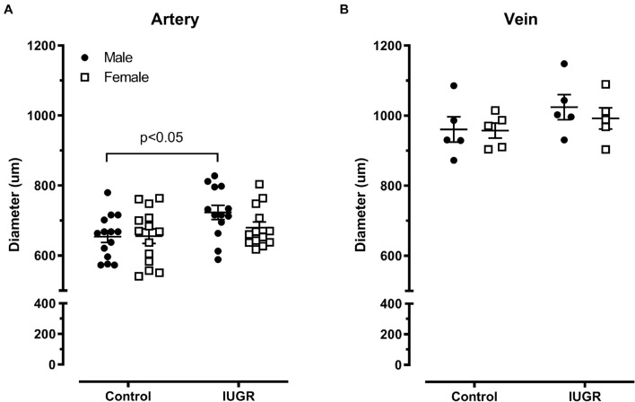
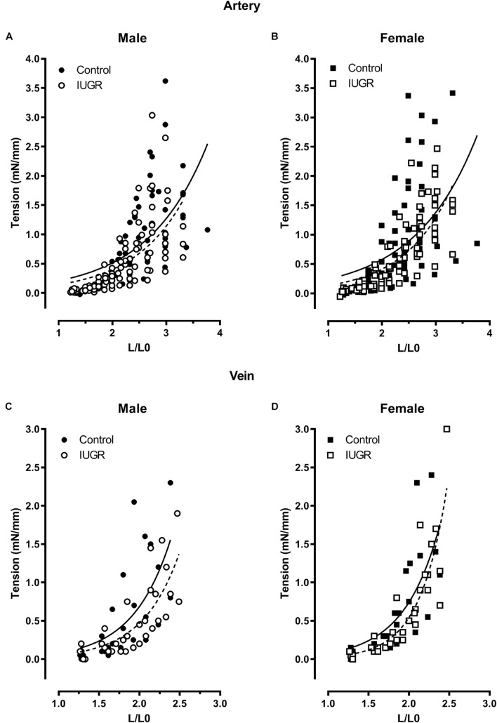
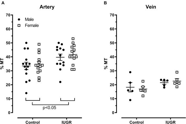
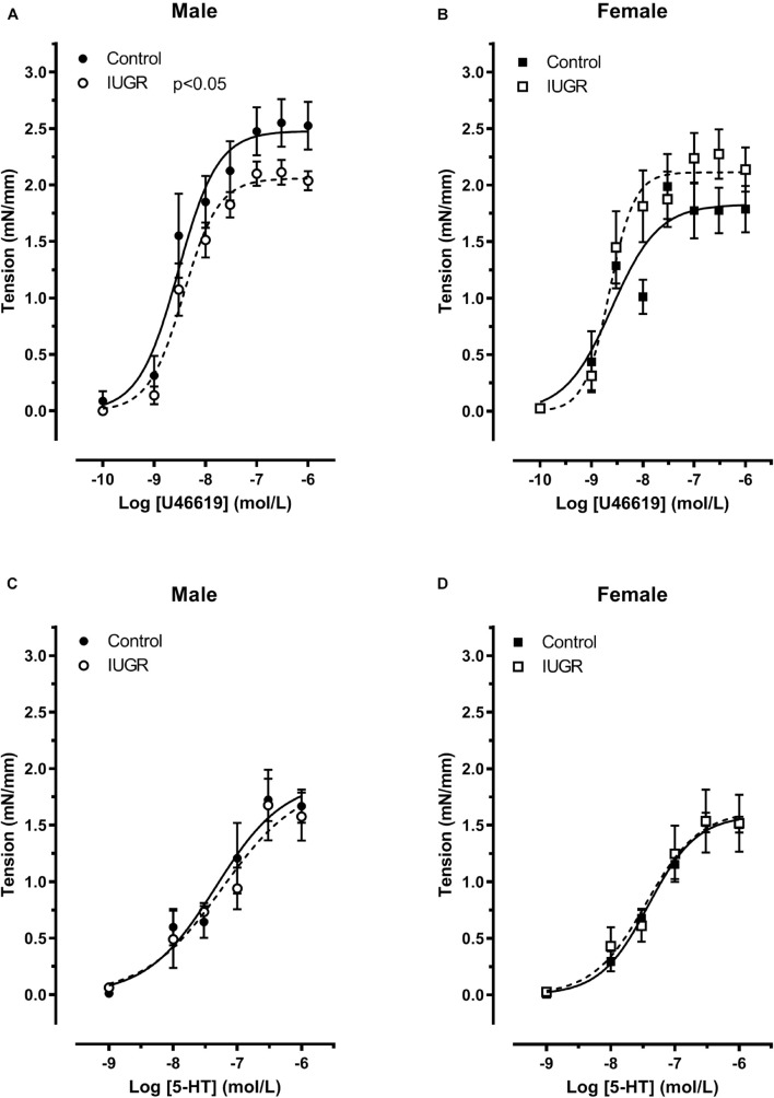
Fetal growth needs adequate blood perfusion from both sides of the placenta, on the maternal side through the uterine vessels and on the fetal side through the umbilical cord. In a model of intrauterine growth restriction (IUGR) induced by reduced blood volume expansion, uterine artery remodeling was blunted. The aim of this study is to determine if IUGR and fetus sex alter the functional and mechanical parameters of umbilical cord blood vessels. Pregnant rats were given a low sodium (IUGR) or a control diet for the last 7 days of pregnancy. Umbilical arteries and veins from term (22 day) fetal rats were isolated and set-up in wire myographs. Myogenic tone, diameter, length tension curve and contractile response to thromboxane analog U46619 and serotonin (5-HT) were measured. In arteries from IUGR fetuses, myogenic tone was increased in both sexes while diameter was significantly greater only in male fetuses. In umbilical arteries collected from the control group, the maximal contraction to U46619 was lower in females than males. Compared to the control groups, the maximal response decreased in IUGR male arteries and increased in female ones, thus abolishing the sexual dimorphism observed in the control groups. Reduced contractile response to U46619 was observed in the IUGR vein of both sexes. No difference between groups was observed in response to 5HT in arteries. In conclusion, the change in parameters of the umbilical cord blood vessels in response to a mild insult seems to show adaptation that favors better exchange of deoxygenated and wasted blood from the fetus to the placenta with increased myogenic tone.

Keywords: fetal adverse environment; rat; thromboxane analog; umbilical cords; vascular function.


Conflict of interest statement

The authors declare that the research was conducted in the absence of any commercial or financial relationships that could be construed as a potential conflict of interest.

Figures

FIGURE 1
FIGURE 1
Measurement of the internal circumference. f, distance in μm between the inner edges of the wires; tg wire, tungsten wire; d, diameter of the wire. Internal circumference (L) is given by: L = (π + 2)d + 2f.
FIGURE 2
FIGURE 2
Determination of optimal internal circumference. Umbilical artery length-tension curve and theoretical straight line of Laplace (for a pressure of 20, 30, 40, and 50 mmHg) (A). Tension developed in response to 60 mM KCl at different internal circumference in arteries n = 8 (B) and veins n = 4 (C). Arrow indicated the optimal internal circumference for both vessels in subsequent experiments.
FIGURE 3
FIGURE 3
Representative tracing of the experimental protocol in an umbilical artery (similar protocol for the vein). Vessels were set at L0, a first length-tension (L-T) curve is recorded to normalize the vessels at L40 or L20 (A). Then, contractility of the preparation is tested with a 60 mM potassium chloride challenge (B). PSS is replaced by fresh PSS and vessels are allowed to rest 60 min before cumulative concentration-response curve to U46619 or Serotonin (C). After washing the contractile agent response with fresh PSS, Ca2+ free PSS (containing 2 mM EGTA) is added. The vessels are returned to L0, second L-T curve is measured with stretch steps of 100 μm (D). L40 or L20 are recorded and diameter in free calcium PSS calculated and recorded. Myogenic tone is calculated using this formula: % MT for artery = (Diameter L40 in Ca2+ free PSS – Diameter L40 in PSS)/Diameter L40 Ca2+ free PSS × 100 (E).
FIGURE 4
FIGURE 4
Representative tracing of the experimental protocol in an umbilical vein for myogenic tone measurement. Length-tension (L-T) curve in PSS (A). Length-tension curve in Ca2+ free PSS (B). Determination of myogenic tone (C).
FIGURE 5
FIGURE 5
Umbilical artery (A) and vein (B) passive diameter in micrometer (μm) for the different groups of animals. Control (n = 14), IUGR (n = 13). Lines represents mean ± SEM.
FIGURE 6
FIGURE 6
Length-tension relationship in the umbilical artery (A,B, control n = 14, IUGR n = 13) and vein (C,D, n = 5, each group). The lines are the best fit of all experimental points (solid: control; dotted: IUGR).
FIGURE 7
FIGURE 7
Percent myogenic tone (%MT) in the umbilical artery (A) and vein (B). Control (n = 14), IUGR (n = 13). Lines represents mean ± SEM.
FIGURE 8
FIGURE 8
Contractile responses to U46619 (A,B, n = 8) and serotonin (5-HT) (C,D, control n = 6, IUGR n = 5) in umbilical artery of control and IUGR fetuses. Each points is the mean ± SEM of n animals for each groups and sexes. The lines represents the best fit of all experimental points.
FIGURE 9
FIGURE 9
Contractile responses to U46619 in umbilical vein of male (A) and female (B) fetuses. Each points is the mean ± SEM of animals (n = 5) for each groups and sexes. The lines represents the best fit of all experimental points.

Similar articles

Cited by

References

    1. Angus J. A., Wright C. E. (2000). Techniques to study the pharmacodynamics of isolated large and small blood vessels. J. Pharmacol. Toxicol. Methods 44 395–407. 10.1016/s1056-8719(00)00121-0 - DOI - PubMed
    1. Arishima K., Yamamoto M., Ueda Y., Kusanagi M., Eguchi Y. (1990). Developmental changes in the umbilical arteries with observation of the effects of indomethacin in fetal rats. Nihon Juigaku Zasshi 52 807–812. 10.1292/jvms1939.52.807 - DOI - PubMed
    1. Balzano F., Bellu E., Basoli V., Dei Giudici S., Santaniello S., Cruciani S., et al. (2019). Lessons from human umbilical cord: gender differences in stem cells from Wharton’s jelly. Eur. J. Obstet. Gynecol. Reprod. Biol. 234 143–148. 10.1016/j.ejogrb.2018.12.028 - DOI - PubMed
    1. Battista M. C., Oligny L. L., St-Louis J., Brochu M. (2002). Intrauterine growth restriction in rats is associated with hypertension and renal dysfunction in adulthood. Am. J. Physiol. Endocrinol. Metab. 283 E124–E131. - PubMed
    1. Baykal C., Sargon M. F., Esinler I., Onderoglu S., Onderoglu L. (2004). Placental microcirculation of intrauterine growth retarded fetuses: scanning electron microscopy of placental vascular casts. Arch. Gynecol. Obstet. 270 99–103. 10.1007/s00404-003-0511-z - DOI - PubMed

LinkOut - more resources