

Neu5Ac Induces Human Dental Pulp Stem Cell Osteo-/Odontoblastic Differentiation by Enhancing MAPK/ERK Pathway Activation
- PMID: 34603453
- PMCID: PMC8483915
- DOI: 10.1155/2021/5560872
Neu5Ac Induces Human Dental Pulp Stem Cell Osteo-/Odontoblastic Differentiation by Enhancing MAPK/ERK Pathway Activation
Abstract
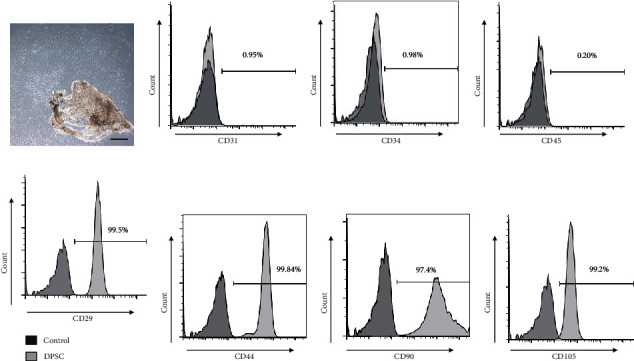
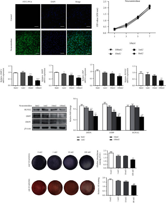
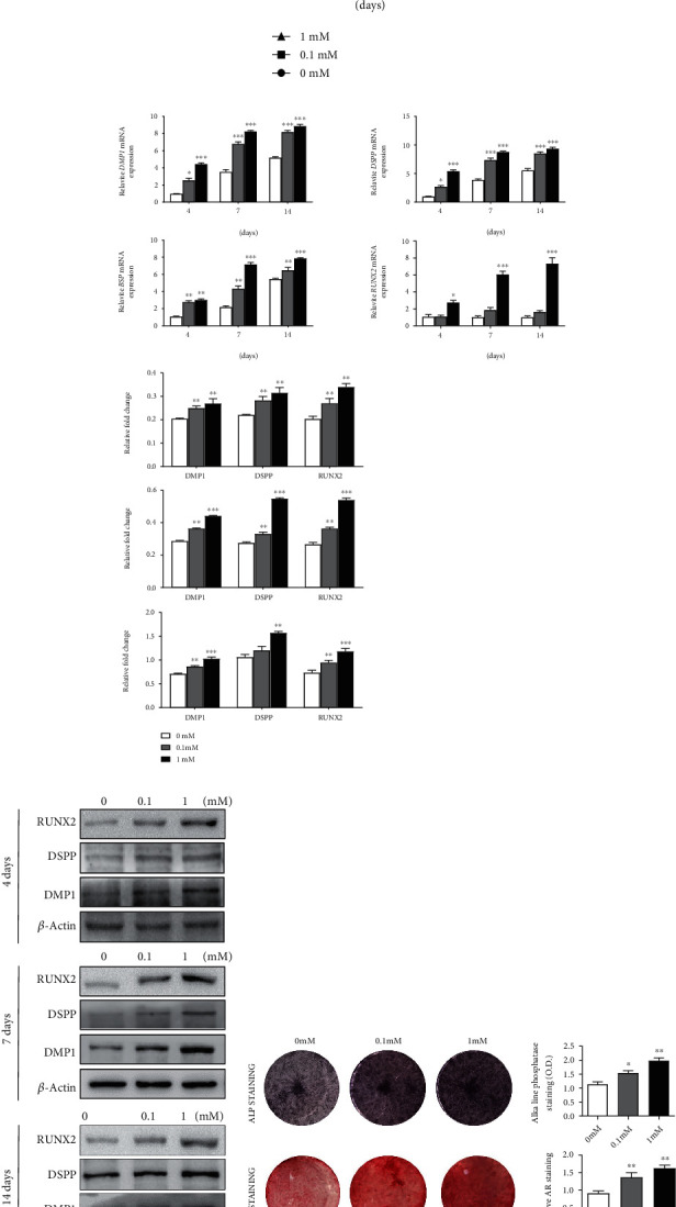
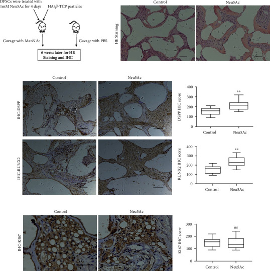
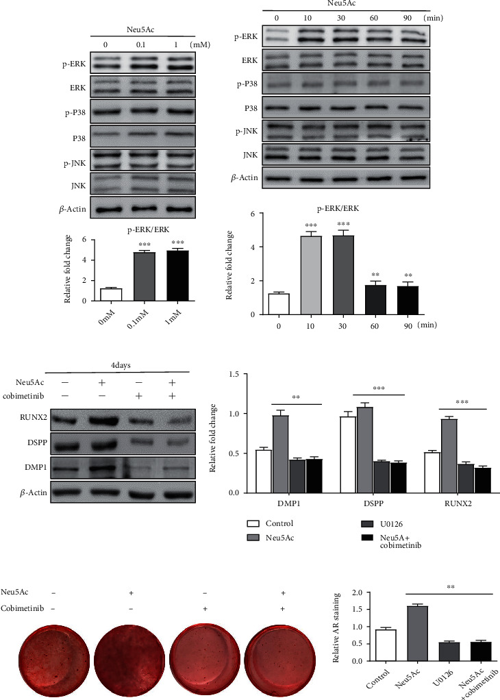
Dental pulp stem cells (DPSCs) must undergo odontoblastic differentiation in order to facilitate the process of dentin-pulp complex repair. Herein, we sought to explore the ability of Neu5Ac (one form of sialic acid) to influence DPSC osteo-/odontoblastic differentiation via modulating mitogen-activated protein kinase (MAPK) signaling. Methodology. DPSCs were isolated from human third permanent teeth and were grown in vitro. Fluorescent microscopy was used to detect the existence of sialic acid on the DPSC membrane. Following the treatment of different concentrations of Neu5Ac and removing sialic acid from the cell surface by neuraminidase, the osteo-/odontoblastic differentiation of these cells was evaluated via mineralization, alkaline phosphatase, and in vivo assays. In addition, the expression of genes related to osteo-/odontoblastic differentiation and MAPK signaling at different stages of this differentiation process was analyzed in the presence or absence of Neu5Ac. Results. The existence of sialic acid on the DPSC membrane was confirmed by fluorescent microscopy, and the ability of osteo-/odontoblastic differentiation was decreased after removing sialic acid by neuraminidase. Treatment of DPSCs with Neu5Ac (0.1 mM or 1 mM) significantly enhanced their mineralization ability and alkaline phosphatase activity. The expression levels of DMP1, DSPP, BSP, and RUNX2 were also increased. Treatment of nude mice with ManNAc (the prerequisite form of Neu5Ac) also enhanced DPSC mineralization activity in vivo. Furthermore, Neu5Ac treatment enhanced p-ERK expression in DPSCs, while ERK pathway inhibition disrupted the ability of Neu5Ac to enhance the osteo-/odontoblastic differentiation of these cells. Conclusions. Neu5Ac can promote DPSC osteo-/odontoblastic differentiation through a process associated with the modulation of the ERK signaling pathway activity.
Copyright © 2021 Changzhou Li et al.
Conflict of interest statement
The authors declare that they have no conflicts of interest.
Figures
Similar articles
-
The odontoblastic differentiation of dental mesenchymal stem cells: molecular regulation mechanism and related genetic syndromes.Front Cell Dev Biol. 2023 Sep 25;11:1174579. doi: 10.3389/fcell.2023.1174579. eCollection 2023. Front Cell Dev Biol. 2023. PMID: 37818127 Free PMC article. Review.
-
Epiregulin enhances odontoblastic differentiation of dental pulp stem cells via activating MAPK signalling pathway.Cell Prolif. 2019 Nov;52(6):e12680. doi: 10.1111/cpr.12680. Epub 2019 Aug 27. Cell Prolif. 2019. PMID: 31454111 Free PMC article.
-
Depletion of EREG enhances the osteo/dentinogenic differentiation ability of dental pulp stem cells via the p38 MAPK and Erk pathways in an inflammatory microenvironment.BMC Oral Health. 2021 Jun 21;21(1):314. doi: 10.1186/s12903-021-01675-0. BMC Oral Health. 2021. PMID: 34154572 Free PMC article.
-
Hyaluronan induces odontoblastic differentiation of dental pulp stem cells via CD44.Stem Cell Res Ther. 2016 Sep 20;7(1):135. doi: 10.1186/s13287-016-0399-8. Stem Cell Res Ther. 2016. PMID: 27651223 Free PMC article.
-
Biological functions of sialic acid as a component of bacterial endotoxin.Front Microbiol. 2022 Oct 20;13:1028796. doi: 10.3389/fmicb.2022.1028796. eCollection 2022. Front Microbiol. 2022. PMID: 36338080 Free PMC article. Review.
Cited by
-
Caffeic Acid Phenethyl Ester Induces Vascular Endothelial Growth Factor Production and Inhibits CXCL10 Production in Human Dental Pulp Cells.Curr Issues Mol Biol. 2022 Nov 15;44(11):5691-5699. doi: 10.3390/cimb44110385. Curr Issues Mol Biol. 2022. PMID: 36421669 Free PMC article.
-
The odontoblastic differentiation of dental mesenchymal stem cells: molecular regulation mechanism and related genetic syndromes.Front Cell Dev Biol. 2023 Sep 25;11:1174579. doi: 10.3389/fcell.2023.1174579. eCollection 2023. Front Cell Dev Biol. 2023. PMID: 37818127 Free PMC article. Review.
-
Potential role of dental pulp stem cells conditioned medium for odontoblastic differentiation.Biol Res. 2022 Mar 4;55(1):11. doi: 10.1186/s40659-022-00380-8. Biol Res. 2022. PMID: 35246266 Free PMC article.
References
LinkOut - more resources
Full Text Sources
Other Literature Sources
Miscellaneous
