Review
. 2021 Sep 22:2021:8502021.
doi: 10.1155/2021/8502021. eCollection 2021.

Design and Optimization of the Circulatory Cell-Driven Drug Delivery Platform

Affiliations
Review

Design and Optimization of the Circulatory Cell-Driven Drug Delivery Platform

Pengyu Gao et al. Stem Cells Int. .

Abstract

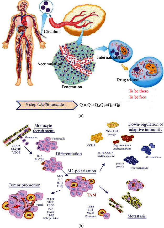
Achievement of high targeting efficiency for a drug delivery system remains a challenge of tumor diagnoses and nonsurgery therapies. Although nanoparticle-based drug delivery systems have made great progress in extending circulation time, improving durability, and controlling drug release, the targeting efficiency remains low. And the development is limited to reducing side effects since overall survival rates are mostly unchanged. Therefore, great efforts have been made to explore cell-driven drug delivery systems in the tumor area. Cells, particularly those in the blood circulatory system, meet most of the demands that the nanoparticle-based delivery systems do not. These cells possess extended circulation times and innate chemomigration ability and can activate an immune response that exerts therapeutic effects. However, new challenges have emerged, such as payloads, cell function change, cargo leakage, and in situ release. Generally, employing cells from the blood circulatory system as cargo carriers has achieved great benefits and paved the way for tumor diagnosis and therapy. This review specifically covers (a) the properties of red blood cells, monocytes, macrophages, neutrophils, natural killer cells, T lymphocytes, and mesenchymal stem cells; (b) the loading strategies to balance cargo amounts and cell function balance; (c) the cascade strategies to improve cell-driven targeting delivery efficiency; and (d) the features and applications of cell membranes, artificial cells, and extracellular vesicles in cancer treatment.


Conflict of interest statement

The authors declare no conflict of interest, financial or otherwise.

Figures

Figure 1
Figure 1
(a) Scheme of the CAPIR cascade of a nanomedicine to deliver a free drug into cancer cells. The overall efficiency, Q, is the product of the efficiencies of five steps. Reproduced with permission [4]. Copyright 2017. John Wiley and Sons. (b) Overview of molecules that can recruit monocytes/macrophages to tumor sites and turn into tumor-associated macrophages. Reproduced with permission [26]. Copyright 2009. John Wiley and Sons.
Figure 2
Figure 2
(a) Scheme of NPs conjugated with cell membranes via electrostatic interaction. (b) Dox was bound on the MSC membranes via the CD90/CD73 antibody-ligand interaction. Reproduced with permission [52]. Copyright 2011. American Chemical Society. (c) FluoSpheres (red) modified by NeutrAvidin (orange) to bind to biotinylated MSC membranes. Reproduced with permission [58]. Copyright 2010. American Chemical Society. (d) SN-38 NPs were anchored on T cells via a thiol group expressed on the cell membrane. Reproduced with permission [12]. Copyright 2015. American Association for the Advancement of Science.
Figure 3
Figure 3
Backpack and encapsulation approaches. (a) Scanning electron microscope (SEM) images of RBCs which were backpacked with multitheranostic probes for cancer surgery guidance and therapy. (b) SEM images of higher magnification of cargo-loading RBCs, and the insert shows naked RBC images. Reproduced with permission [48]. Copyright 2019. Ivyspring International Publisher. (c) Confocal micrographs of quantum dot distribution in MSCs after 15–30 min, 1 h, 6 h, and 24–48 h incubation. Nuclei, Hoechst blue; actin, Phalloidin green; quantum dots, red. Scale bar for main images, 15 μm; scale bar for insert images, 10 μm. Reproduced with permission [40]. Copyright 2017. Dove Medical Press.
Figure 4
Figure 4
Three drug release patterns. (a) Exocytosis: either free Dox or Dox/SiO2 NPs were secreted by MAs for glioblastoma treatment. Reproduced with permission [18]. Copyright 2018. John Wiley and Sons. (b) Cell disintegration I: Abraxane-encapsulated NEs disintegrated and formed the neutrophil extracellular traps to kill gastric cancer. Reproduced with permission [38]. Copyright 2018. John Wiley and Sons. (c) Cell disintegration II: Dox-encapsulated and Ce6-backpacked RBCs were disintegrated by photoradiation for breast cancer. Reproduced with permission [46]. Copyright 2017. American Chemical Society. (d) Cell-drug dissociation: SN-38 was dissociated from the T cell membrane into lymphoma cells. Reproduced with permission [12]. Copyright 2015. American Association for the Advancement of Science.
Figure 5
Figure 5
(a) Scheme of gene/chemotherapy on a blood exosome basis. Chemotherapeutic drugs and the cholesterol-modified miRNA21 inhibitor were embedded between the vesicle lipid bilayers for tumor killing; both magnetic molecules and L17E peptides were bound onto the vesicle membrane, respectively, for targeting and lysosome escape. (b) According to nanoparticle tracking analysis (NTA), the original exosome size was 93 nm. (c) The size was increased to 106 nm on average, after cargo loading and membrane modification, and transmission electron microscope (TEM) images showed modified exosomes that retained a clustered structure. Reproduced with permission [90]. Copyright 2020. Ivyspring International Publisher.

Similar articles

Cited by

References

    1. World Health Organization. WHO - the top 10 causes of death. May 2018, http://www.who.int/en/news-room/fact-sheets/detail/the-top-10-causes-of-....
    1. Shi J., Kantoff P. W., Wooster R., Farokhzad O. C. Cancer nanomedicine: progress, challenges and opportunities. Nature Reviews Cancer . 2017;17(1):20–37. doi: 10.1038/nrc.2016.108. - DOI - PMC - PubMed
    1. Jing L., Hao H., Peng M., Yuanquan W. Current status and prospect of cancer nanomedicine in clinical translation. Science & Technology Review . 2018;36(22):118–126.
    1. Sun Q., Zhou Z., Qiu N., Shen Y. Rational design of cancer nanomedicine: nanoproperty integration and synchronization. Advanced Materials . 2017;29(14):1–18. doi: 10.1002/adma.201606628. - DOI - PubMed
    1. Betker J. L., Jones D., Childs C. R., et al. Nanoparticle uptake by circulating leukocytes: a major barrier to tumor delivery. Journal of Controlled Release . 2018;286:85–93. doi: 10.1016/j.jconrel.2018.07.031. - DOI - PMC - PubMed

LinkOut - more resources