

Etomidate inhibits cell proliferation and induces apoptosis in A549 non-small cell lung cancer cells via downregulating WWP2
- PMID: 34603522
- PMCID: PMC8453325
- DOI: 10.3892/etm.2021.10689
Etomidate inhibits cell proliferation and induces apoptosis in A549 non-small cell lung cancer cells via downregulating WWP2
Abstract
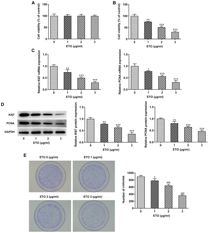
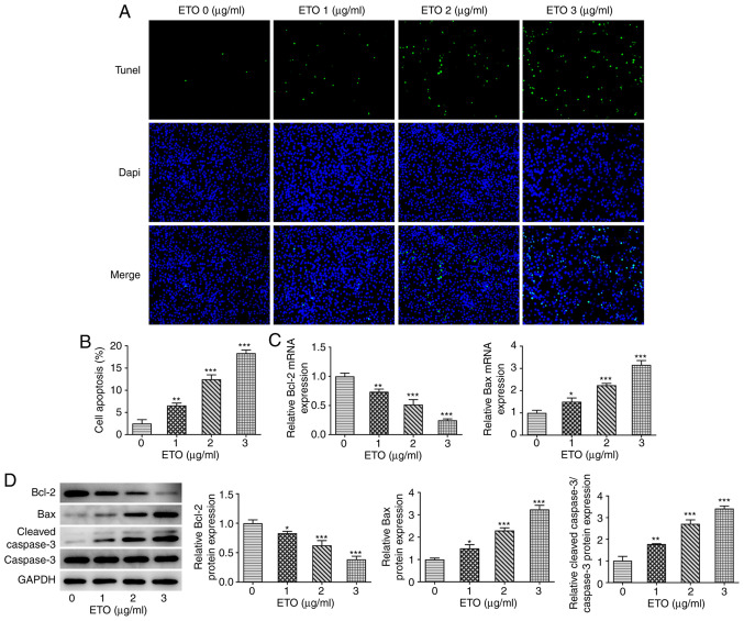
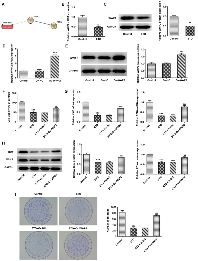
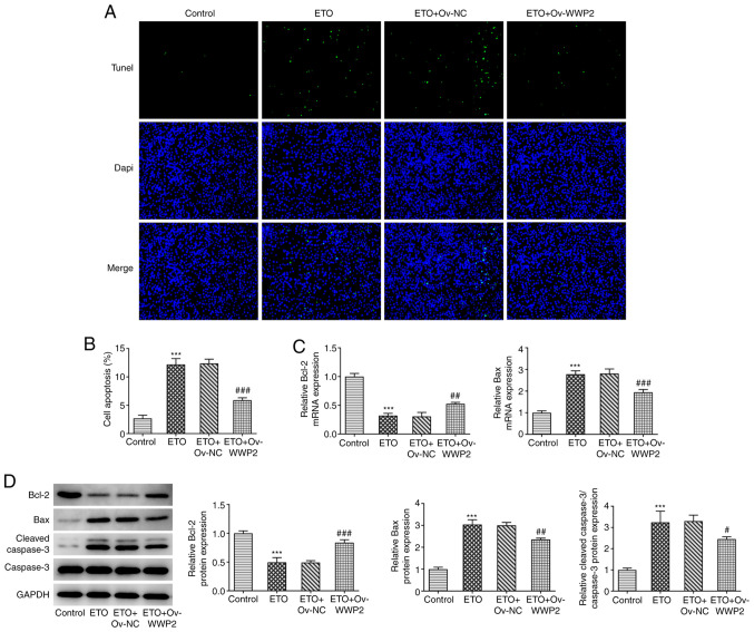
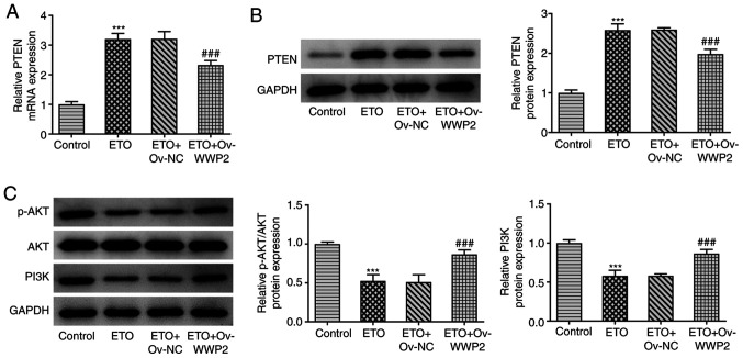
Etomidate (ETO) is a commonly used intravenous anesthetic that has been reported to exert a tumor suppressive effect in several types of cancer. The present study aimed to investigate the effect of ETO on cell proliferation and apoptosis in non-small cell lung cancer (NSCLC) cells and elucidate its potential mechanism of action. Therefore, Cell Counting Kit-8 assay was performed to evaluate the effect of different concentrations of ETO (0, 1, 2 or 3 µg/ml) on A549 cell viability. In addition, the possible interaction between ETO and WW domain containing E3 ubiquitin protein ligase 2 (WWP2) was predicted using the STITCH database. Additionally, a stable WWP2-overexpressing A549 cell line was constructed by transfecting A549 cells with the pcDNA3.1-WWP2 plasmid. Cell proliferation and apoptosis were assessed using colony formation and TUNEL assays, respectively. The mRNA and protein expression levels of the apoptosis-related proteins Bcl-2, Bax, caspase 3 and cleaved-caspase 3 were determined by reverse transcription-quantitative PCR and western blotting. In addition, the expression and phosphorylation levels of proliferation-associated genes (PCNA and Ki-67) and proteins in the PI3K/Akt pathway were analyzed by western blotting. The results showed that treatment with ETO attenuated the cell viability and proliferation of A549 cells. ETO also promoted cell apoptosis and decreased the expression of the anti-apoptotic protein Bcl-2, whilst increasing that of pro-apoptotic proteins Bax and cleaved caspase 3 in a dose-dependent manner. Furthermore, ETO was found to negatively regulate the expression of WWP2, such that WWP2 overexpression reversed the potentiating effects of ETO on cell apoptosis. In addition, ETO promoted the expression of PTEN and reduced the phosphorylation levels of the PI3K/AKT pathway-related proteins. These effects aforementioned could also be reversed by WWP2 overexpression. Therefore, data from the present study suggest that ETO can attenuate the progression of NSCLC through by the PI3K/AKT pathway, specifically by targeting WWP2. These findings may provide a novel target for the treatment of NSCLC.
Keywords: WW domain-containing E3 ubiquitin protein ligase 2; apoptosis; etomidate; non-small cell lung cancer; proliferation.
Copyright: © Li et al.
Conflict of interest statement
The authors declare that they have no competing interests.
Figures
Similar articles
-
WWP2 binds to NKRF, enhances the NF-κB signaling, and promotes malignant phenotypes of acute myeloid leukemia cells.Biochem Cell Biol. 2024 Feb 1;102(1):85-95. doi: 10.1139/bcb-2022-0360. Epub 2023 Nov 3. Biochem Cell Biol. 2024. PMID: 37921219
-
Inhibition of WWP2 suppresses proliferation, and induces G1 cell cycle arrest and apoptosis in liver cancer cells.Mol Med Rep. 2016 Mar;13(3):2261-6. doi: 10.3892/mmr.2016.4771. Epub 2016 Jan 13. Mol Med Rep. 2016. PMID: 26783238
-
ARHGAP24 inhibits cell proliferation and cell cycle progression and induces apoptosis of lung cancer via a STAT6-WWP2-p27 axis.Carcinogenesis. 2020 Jul 10;41(5):711-721. doi: 10.1093/carcin/bgz144. Carcinogenesis. 2020. PMID: 31430374 Free PMC article.
-
Apoptosis induced by the methanol extract of Salvia miltiorrhiza Bunge in non-small cell lung cancer through PTEN-mediated inhibition of PI3K/Akt pathway.J Ethnopharmacol. 2017 Mar 22;200:107-116. doi: 10.1016/j.jep.2016.12.051. Epub 2017 Jan 12. J Ethnopharmacol. 2017. PMID: 28088493
-
miR‑379‑5p inhibits cell proliferation and promotes cell apoptosis in non‑small cell lung cancer by targeting β‑arrestin‑1.Mol Med Rep. 2020 Dec;22(6):4499-4508. doi: 10.3892/mmr.2020.11553. Epub 2020 Sep 30. Mol Med Rep. 2020. PMID: 33173959 Free PMC article.
Cited by
-
Molecular authentication, metabolite profiling and in silico-in vitro cytotoxicity screening of endophytic Penicillium ramusculum from Withania somnifera for breast cancer therapeutics.3 Biotech. 2024 Mar;14(3):64. doi: 10.1007/s13205-023-03906-3. Epub 2024 Feb 9. 3 Biotech. 2024. PMID: 38344285 Free PMC article.
-
WWP2 drives the progression of gastric cancer by facilitating the ubiquitination and degradation of LATS1 protein.Cell Commun Signal. 2023 Feb 17;21(1):38. doi: 10.1186/s12964-023-01050-2. Cell Commun Signal. 2023. PMID: 36803368 Free PMC article.
-
Advances of E3 ligases in lung cancer.Biochem Biophys Rep. 2024 May 27;38:101740. doi: 10.1016/j.bbrep.2024.101740. eCollection 2024 Jul. Biochem Biophys Rep. 2024. PMID: 38841185 Free PMC article. Review.
-
Half the Chromosome It Used to Be: Identifying Cancer Treatments Targeting Aneuploid Losses.Genes (Basel). 2025 Jun 14;16(6):708. doi: 10.3390/genes16060708. Genes (Basel). 2025. PMID: 40565600 Free PMC article.
-
Effects of perioperative anesthetics on the postoperative prognosis of patients undergoing surgery for cervical cancer.Front Pharmacol. 2025 Mar 10;16:1536663. doi: 10.3389/fphar.2025.1536663. eCollection 2025. Front Pharmacol. 2025. PMID: 40129947 Free PMC article. Review.
References
LinkOut - more resources
Full Text Sources
Research Materials
Miscellaneous