Review
. 2021 Nov;22(5):1264.
doi: 10.3892/etm.2021.10699. Epub 2021 Sep 6.

Current diagnosis and treatment of rhinosinusal aspergilloma (Review)

Affiliations
Review

Current diagnosis and treatment of rhinosinusal aspergilloma (Review)

Daniela Vrinceanu et al. Exp Ther Med. 2021 Nov.

Abstract

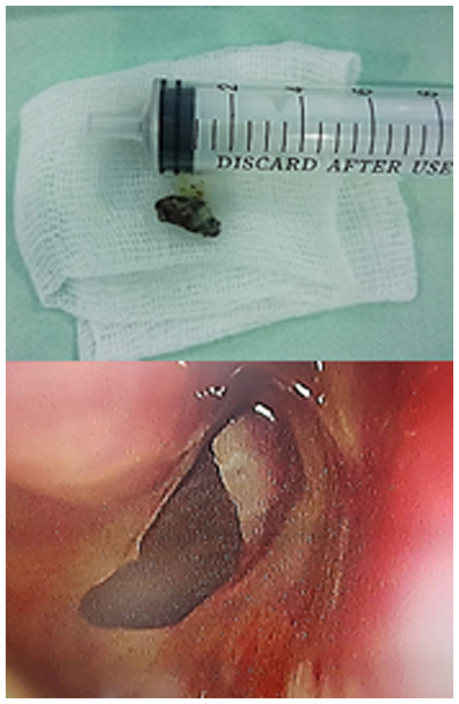
There are numerous types of sinusitis caused by fungal strains, some of which already colonize the nasal cavity. Mild forms present fungus balls growing inside a preexisting sinus cavity. The invasive type ranges from chronic manifestations to acute aggravated episodes. The latter scenario is encountered in cases with reduced immune responses, such as patients with diabetes, individuals receiving any form of transplant, AIDS cases and chemotherapy patients. Without the control of immunosuppression, the infection is aggravated and extends to the orbit and inside the skull base, regardless of the prompt surgical and medical treatment. This is the most common pathogenic fungus on the nasal sinuses level. It can occasionally enter the sinus cavity during dental procedures. The pathogenesis is enhanced by anaerobic conditions in poorly ventilated sinus cavities. Rhinosinusal aspergilloma has a slow, insidious evolution over months and even years. Our experience revealed the presence of both a dental problem and previous self-administered antibiotic regimens in almost every case. The initial symptoms are common with sinusitis of dental origin, but aspergilloma should be considered when a patient with a competent immune system does not respond to standard antibiotic treatment. The final diagnosis of rhinosinusal aspergilloma is conducted on a pathology sample with silver staining. The bacteriology exam of the sinus secretion rarely reveals a fungus infection; however, as revealed in our clinical experience, there may be coinfection with other multidrug-resistant bacteria. Surgical treatment must establish a wide exposure of the sinus cavity and correct drainage regardless of the external, combined or endoscopic approach. Early diagnosis and emergency surgical debridement along with administering systemic antifungal compounds in some cases represent the key to the successful treatment of invasive aspergilloma.

Keywords: aspergilloma; diagnosis; pathology; rhinosinusal; treatment.


Conflict of interest statement

The authors declare that they have no competing interests.

Figures

Figure 1
Figure 1
Coronal CT scan revealing the appearance of fungal right rhinosinusitis with full right maxillary sinus, with obstruction of the osteo-meatal complex, lysis of the medial wall, of the middle nasal cornet skeleton, partially of the bony septum and with suggestive image of aspergilloma in the right middle meatus. CT, computed tomography.
Figure 2
Figure 2
Hyphae and ‘fruiting’ (conidial) heads with non-invasive aspects. Hematoxylin and eosin staining. Magnification, x400.
Figure 3
Figure 3
Classic image of the aspergilloma extracted endoscopically from the right sphenoid sinus and final surgical image with normal mucosa aspect.
Figure 4
Figure 4
Surgical aspect with necrotic maxillary bone and gelatinous modified content of the mucosa in an invasive form of aspergilloma.
Figure 5
Figure 5
Endoscopic image of the aspergilloma situated in the sphenoid ostium along with puss secretion.
Figure 6
Figure 6
Aspergilloma in the right maxillary sinus with the development of a bony septum and evolution of the fungal infection in the lateral compartment.
Figure 7
Figure 7
Dental panoramic X-ray with apical granuloma on the same side with the right sinus pathology.
Figure 8
Figure 8
Clinical aspect of invasive right maxillary sinus aspergilloma in an immunocompromised host with complicated right orbit cellulitis.
Figure 9
Figure 9
Endoscopic image depicting the removal of a right middle meatus aspergilloma.
Figure 10
Figure 10
Surgical aspect of a giant aspergilloma occupying the entire sinus cavity.
Figure 11
Figure 11
Surgical sample showing aspergilloma plus adjacent modified mucosa.
Figure 12
Figure 12
Fungus ball characteristic of Aspergillus sp. Hematoxylin and eosin staining. Magnification, x200.

Similar articles

Cited by

References

    1. Neville BW, Damm DD, Allen CM, Bouquot JE. Fungal and protozoal diseases. In: Oral and Maxillofacial Pathology. 2nd edition. Saunders, Philadelphia PA, pp189-211, 2005.
    1. Lin SJ, Schranz J, Teutsch SM. Aspergillosis case-fatality rate: Systematic review of the literature. Clin Infect Dis. 2001;32:358–366. doi: 10.1086/318483. - DOI - PubMed
    1. Nikolaizik WH, Weichel M, Blaser K, Crameri R. Intracutaneous tests with recombinant allergens in cystic fibrosis patients with allergic bronchopulmonary aspergillosis and aspergillus allergy. Am J Respir Crit Care Med. 2002;165:916–921. doi: 10.1164/ajrccm.165.7.2109008. - DOI - PubMed
    1. Hartl D, Latzin P, Zissel G, Krane M, Krauss-Etschmann S, Griese M. Chemokines indicate allergic bronchopulmonary aspergillosis in patients with cystic fibrosis. Am J Respir Crit Care Med. 2006;173:1370–1376. doi: 10.1164/rccm.200508-1271OC. - DOI - PubMed
    1. Kraemer R, Deloséa N, Ballinari P, Gallati S, Crameri R. Effect of allergic bronchopulmonary aspergillosis on lung function in children with cystic fibrosis. Am J Respir Crit Care Med. 2006;174:1211–1220. doi: 10.1164/rccm.200603-423OC. - DOI - PubMed