. 2021 Sep 23:2021:9925771.
doi: 10.1155/2021/9925771. eCollection 2021.

Hydrogen Sulfide Attenuates Angiotensin II-Induced Cardiac Fibroblast Proliferation and Transverse Aortic Constriction-Induced Myocardial Fibrosis through Oxidative Stress Inhibition via Sirtuin 3

Affiliations

Hydrogen Sulfide Attenuates Angiotensin II-Induced Cardiac Fibroblast Proliferation and Transverse Aortic Constriction-Induced Myocardial Fibrosis through Oxidative Stress Inhibition via Sirtuin 3

Lulu Liu et al. Oxid Med Cell Longev. .

Abstract

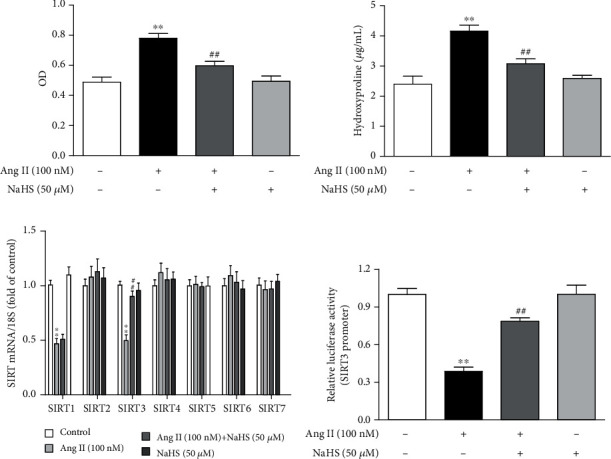
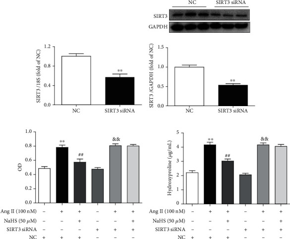
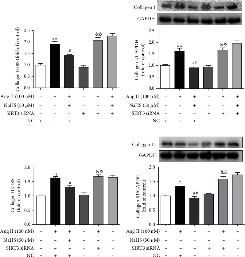
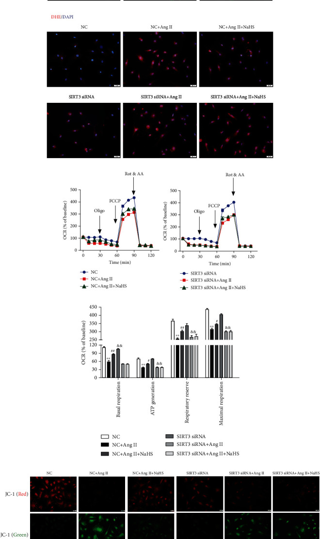
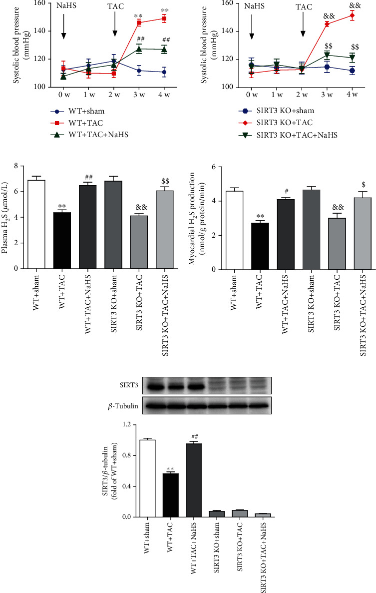
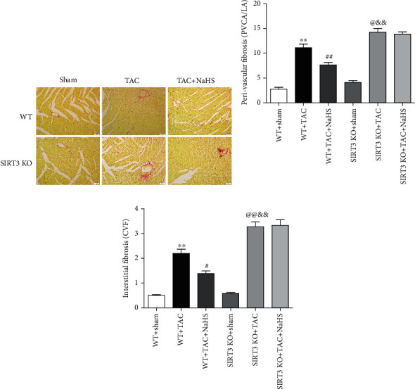
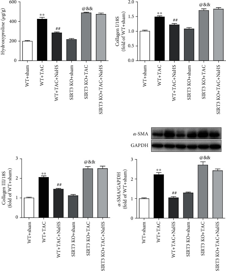
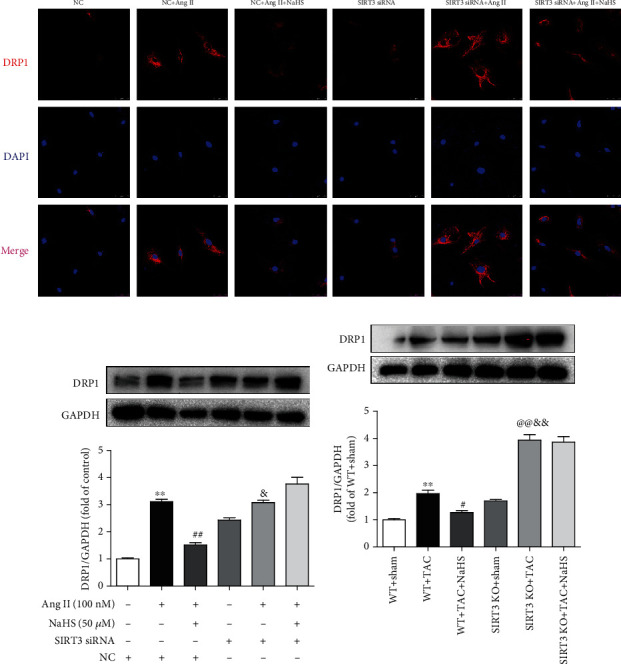
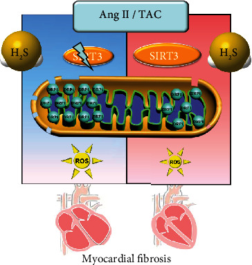
Sirtuin 3 (SIRT3) is critical in mitochondrial function and oxidative stress. Our present study investigates whether hydrogen sulfide (H2S) attenuated myocardial fibrosis and explores the possible role of SIRT3 on the protective effects. Neonatal rat cardiac fibroblasts were pretreated with NaHS followed by angiotensin II (Ang II) stimulation. SIRT3 was knocked down with siRNA technology. SIRT3 promoter activity and expression, as well as mitochondrial function, were measured. Male wild-type (WT) and SIRT3 knockout (KO) mice were intraperitoneally injected with NaHS followed by transverse aortic constriction (TAC). Myocardium sections were stained with Sirius red. Hydroxyproline content, collagen I and collagen III, α-smooth muscle actin (α-SMA), and dynamin-related protein 1 (DRP1) expression were measured both in vitro and in vivo. We found that NaHS enhanced SIRT3 promoter activity and increased SIRT3 mRNA expression. NaHS inhibited cell proliferation and hydroxyproline secretion, decreased collagen I, collagen III, α-SMA, and DRP1 expression, alleviated oxidative stress, and improved mitochondrial respiration function and membrane potential in Ang II-stimulated cardiac fibroblasts, which were unavailable after SIRT3 was silenced. In vivo, NaHS reduced hydroxyproline content, ameliorated perivascular and interstitial collagen deposition, and inhibited collagen I, collagen III, and DRP1 expression in the myocardium of WT mice but not SIRT3 KO mice with TAC. Altogether, NaHS attenuated myocardial fibrosis through oxidative stress inhibition via a SIRT3-dependent manner.


Conflict of interest statement

The authors declare that there is no conflict of interest regarding the publication of this paper.

Figures

Figure 1
Figure 1
NaHS inhibits cell proliferation but enhances SIRT3 transcription in Ang II-stimulated cardiac fibroblasts. After pretreatment with NaHS (50 μM) for 4 h, neonatal rat cardiac fibroblasts were stimulated by Ang II (100 nM) for 24 h. (a) The number of cardiac fibroblasts was detected with CCK-8. (b) The content of hydroxyproline in the cell culture medium was measured. (c) Expression of SIR2 family (SIRT1-7) mRNA was measured by real-time PCR. (d) After the SIRT3 promoter luciferase plasmid was transfected into cardiac fibroblasts for 24 h, cells were pretreated with NaHS (50 μM) for 4 h followed by Ang II (100 nM) stimulation for another 24 h. Relative promoter activity of SIRT3 was detected with a dual-luciferase reporter assay system. ∗∗P < 0.01 as compared with untreated cells; ##P < 0.01 as compared with Ang II alone-stimulated cells. n = 6.
Figure 2
Figure 2
NaHS inhibits Ang II-stimulated cardiac fibroblast proliferation via SIRT3. (a, b) After SIRT3 siRNA or NC siRNA was transfected into neonatal rat myocardial fibroblasts for 48 h, expression of SIRT3 mRNA and protein was measured by real-time PCR and western blot, respectively. ∗∗P < 0.01 as compared with cells with NC siRNA transfection. n = 6. (c) After SIRT3 siRNA or NC siRNA was transfected into cardiac fibroblasts for 24 h, the cells were pretreated with NaHS (50 μM) for 4 h followed by Ang II (100 nM) stimulation for another 24 h. The number of cardiac fibroblasts was detected with CCK-8. (d) The content of hydroxyproline in the cell culture medium was measured. ∗∗P < 0.01 as compared with untreated cells with NC siRNA transfection; ##P < 0.01 as compared with Ang II alone-stimulated cells with NC siRNA transfection; &&P < 0.01 as compared with untreated cells with SIRT3 siRNA transfection. n = 6.
Figure 3
Figure 3
NaHS suppresses collagen expression in Ang II-stimulated cardiac fibroblasts via SIRT3. After SIRT3 siRNA or NC siRNA was transfected into neonatal rat cardiac fibroblasts for 24 h, the cells were pretreated with NaHS (50 μM) for 4 h followed by Ang II (100 nM) stimulation for another 24 h. (a, b) Expression of collagen I mRNA and protein was measured by real-time PCR and western blot, respectively. (c, d) Expression of collagen III mRNA and protein was measured by real-time PCR and western blot, respectively. P < 0.05 and ∗∗P < 0.01 as compared with untreated cells with NC siRNA transfection; #P < 0.05 and ##P < 0.01 as compared with Ang II alone-stimulated cells with NC siRNA transfection; &&P < 0.01 as compared with untreated cells with SIRT3 siRNA transfection. n = 6.
Figure 4
Figure 4
NaHS inhibits α-SMA expression and oxidative stress and improves mitochondrial respiration function and membrane potential in Ang II-stimulated cardiac fibroblasts via SIRT3. After SIRT3 siRNA or NC siRNA was transfected into neonatal rat cardiac fibroblasts for 24 h, the cells were pretreated with NaHS (50 μM) for 4 h followed by Ang II (100 nM) stimulation for another 24 h. (a) Expression of α-SMA in cardiac fibroblasts was measured by immunofluorescence with Alexa Fluor 488 (green)-conjugated IgG. The nuclei were stained using DAPI (blue). Bar = 50 μm. (b) ROS was detected with DHE staining. Bar = 50 μm. (c) The mitochondrial respiration function of cardiac fibroblasts was measured. (d) Quantitative analysis of basal respiration, ATP generation, respiratory reserve capacity, and maximal respiratory. ∗∗P < 0.01 as compared with untreated cells with NC siRNA transfection; #P < 0.05 and ##P < 0.01 as compared with Ang II alone-stimulated cells with NC siRNA transfection; &&P < 0.01 as compared with untreated cells with SIRT3 siRNA transfection. n = 6. (e) Mitochondrial permeability potential was determined by JC-1 staining. Bar = 200 μm.
Figure 5
Figure 5
NaHS decreases blood pressure but restores H2S levels and SIRT3 expression in mice with TAC. After intraperitoneal injection by NaHS (50 μmol·kg−1·day−1) or normal saline (NS) for 2 weeks, male wild-type (WT) mice and SIRT3 knockout (SIRT3 KO) mice were subjected to transverse aortic constriction (TAC) surgery. NaHS or NS was administrated for another 2 weeks. (a) The level of SBP in WT mice and SIRT3 KO mice was monitored every week after NaHS administration. (b) H2S concentration in the plasma was measured. (c) H2S production in the myocardium was detected. (d) Expression of SIRT3 protein in the myocardium was measured by western blot. ∗∗P < 0.01 as compared with WT+Sham; #P < 0.05 and ##P < 0.01 as compared with WT+TAC; &&P < 0.01 as compared with SIRT3 KO+Sham; $$P < 0.01 as compared with SIRT3 KO+TAC. n = 6.
Figure 6
Figure 6
NaHS ameliorates collagen deposition in the myocardium of WT mice but not SIRT3 KO mice with TAC. After intraperitoneal injection by NaHS (50 μmol·kg−1·day−1) or NS for 2 weeks, male WT mice and SIRT3 KO mice were subjected to TAC surgery. NaHS or NS was administrated for another 2 weeks. (a) Collagen deposition in the myocardium was stained with saturated picric acid-Sirius red. Bar = 50 μm. (b) Perivascular fibrosis of the myocardium was assessed by the ratio of the perivascular collagen area (PVCA) to the luminal area (LA). (c) Interstitial fibrosis of the myocardium was assessed by the collagen volume fraction (CVF). ∗∗P < 0.01 as compared with WT+Sham; #P < 0.05 or @P < 0.05 and ##P < 0.01 or @@P < 0.01 as compared with WT+TAC; &&P < 0.01 as compared with SIRT3 KO+Sham. n = 6.
Figure 7
Figure 7
NaHS reduces collagen and α-SMA expressions in the myocardium of WT mice but not SIRT3 KO mice with TAC. After intraperitoneal injection by NaHS (50 μmol·kg−1·day−1) or NS for 2 weeks, male WT mice and SIRT3 KO mice were subjected to TAC surgery. NaHS or NS was administrated for another 2 weeks. (a) The content of hydroxyproline in the myocardium was measured. (b, c) Expression of collagen I and collagen III mRNA in the myocardium was measured by real-time PCR. (d) Expression of α-SMA protein in the myocardium was measured by western blot. ∗∗P < 0.01 as compared with WT+Sham; ##P < 0.01 or @P < 0.05 as compared with WT+TAC; &&P < 0.01 as compared with SIRT3 KO+Sham. n = 8.
Figure 8
Figure 8
NaHS restores DRP1 expression in the cardiac fibroblasts with Ang II stimulation and in the myocardium of mice with TAC via SIRT3. (a) After SIRT3 siRNA or NC siRNA was transfected into neonatal rat cardiac fibroblasts for 24 h, the cells were pretreated with NaHS (50 μM) for 4 h followed by Ang II (100 nM) stimulation for another 24 h. DRP1 expression in cardiac fibroblasts was detected by immunofluorescence with Cy3 (red)-conjugated IgG. The nuclei were stained using DAPI (blue). Bar = 25 μm. (b) Expression of DRP1 protein was measured by western blot. ∗∗P < 0.01 as compared with untreated cells with NC siRNA transfection; ##P < 0.01 as compared with Ang II alone-stimulated cells with NC siRNA transfection; &P < 0.05 as compared with untreated cells with SIRT3 siRNA transfection. n = 6. (c) After intraperitoneal injection by NaHS (50 μmol·kg−1·day−1) or NS for 2 weeks, male WT mice and SIRT3 KO mice were subjected to TAC surgery. NaHS or NS was administrated for another 2 weeks. Expression of DRP1 protein in the myocardium was measured by western blot. ∗∗P < 0.01 as compared with WT+Sham; #P < 0.05 or @@P < 0.01 as compared with WT+TAC; &&P < 0.01 as compared with SIRT3 KO+Sham. n = 6.
Figure 9
Figure 9
Illustration of the mechanism of protective effects on myocardial fibrosis by H2S. H2S enhanced SIRT3 transcription, decreased the DRP1 level, ameliorated mitochondrial membrane rupture, suppressed oxidative stress, and alleviated Ang II-induced cardiac fibroblast proliferation and TAC-induced myocardial fibrosis. However, these protective effects of H2S were unavailable if SIRT3 was silenced in cells or deficient in mice. It suggested that H2S attenuated myocardial fibrosis through oxidative stress inhibition via a SIRT3-dependent manner.

Similar articles

Cited by

References

    1. Frangogiannis N. G. Cardiac fibrosis: cell biological mechanisms, molecular pathways and therapeutic opportunities. Molecular Aspects of Medicine . 2019;65:70–99. doi: 10.1016/j.mam.2018.07.001. - DOI - PubMed
    1. Gonzalez A., Schelbert E. B., Diez J., Butler J. Myocardial interstitial fibrosis in heart failure: biological and translational perspectives. Journal of the American College of Cardiology . 2018;71(15):1696–1706. doi: 10.1016/j.jacc.2018.02.021. - DOI - PubMed
    1. Wang P., Luo L., Shen Q., et al. Rosuvastatin improves myocardial hypertrophy after hemodynamic pressure overload via regulating the crosstalk of Nrf2/ARE and TGF-β/ smads pathways in rat heart. European Journal of Pharmacology . 2018;820:173–182. doi: 10.1016/j.ejphar.2017.12.013. - DOI - PubMed
    1. Song Q., Liu L., Yu J., et al. Dihydromyricetin attenuated Ang II induced cardiac fibroblasts proliferation related to inhibitory of oxidative stress. European Journal of Pharmacology . 2017;807:159–167. doi: 10.1016/j.ejphar.2017.04.014. - DOI - PubMed
    1. Xu F., Sun S., Wang X., Ni E., Zhao L., Zhu W. GRK2 mediates arginine vasopressin-induced interleukin-6 production via nuclear Factor-κB signaling neonatal rat cardiac fibroblast. Molecular Pharmacology . 2017;92(3):278–284. doi: 10.1124/mol.116.107698. - DOI - PubMed

MeSH terms