

Airways cephalometric norms from a sample of Caucasian Children
- PMID: 34603624
- PMCID: PMC8464393
- DOI: 10.4317/jced.58105
Airways cephalometric norms from a sample of Caucasian Children
Abstract
Background: The diagnosis of the respiratory pattern and the analysis of airway dimension using lateral cephalometric radiographs include the study of the adenoid region, free air space of the nasopharynx and oropharynx, soft palate and posterior part of the tongue. The objective of this study is to identify the airways cephalometric norms from a sample of Caucasian children, in relation to gender, age and type of malocclusion.
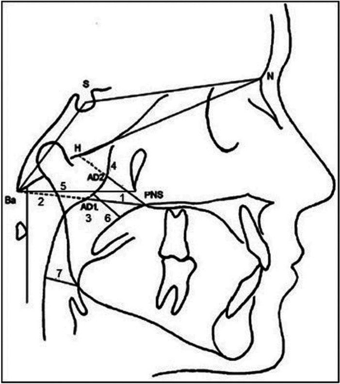
Material and methods: A total of 480 patients of both sexes were included in the study, the age ranged between 6-12 years. The radiographic records were analyzed using the Nemoceph® 11.3.0 software and the diagnosis of skeletal class was performed using the Steiner analysis. The cephalometric measurements used for the study were PNS-AD1, AD1-Ba, PNS-Ba, Ptm-Ba, PNS-H and the upper and lower airways according to McNamara analysis. The comparative analysis was performed using only upper and lower airways variables.
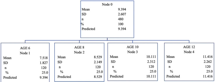
Results: The mean values for each variable in the total sample were 23.2 mm (PNS-Ad1), 24.7 mm (Ad1-Ba), 47.6 mm (PNS-Ba), 45.7 mm (Ptm-Ba), 30.0 mm (PNS-H), 9.3 mm (upper airway) and 11.5 mm (lower airway). According to gender, all variables were greater in the boys group except for the lower airway. In relation to age, the mean values increased with age except for the lower airway and the AD1-Ba variables. In patients with skeletal Class I greater dimensions of the upper and lower airways were observed.
Conclusions: In this Caucasian sample, it has been observed a tendency of minor airway dimensions in patients with skeletal Class II, lower age range female gender. It has been observed only significant differences between age and skeletal class for lower airways variable and, in relation to upper airways variable the results were significant in relation to age. Key words:Child development, Diagnostic XRay, Cephalometry, Respiratory system diagnostic imaging.
Copyright: © 2021 Medicina Oral S.L.
Conflict of interest statement
Conflicts of interest The authors declare no conflict of interest in this article.
Figures
Similar articles
-
Correlation between skeletal changes by maxillary protraction and upper airway dimensions.Angle Orthod. 2011 May;81(3):426-32. doi: 10.2319/082610-499.1. Epub 2011 Feb 7. Angle Orthod. 2011. PMID: 21299388 Free PMC article.
-
A Comparative Assessment of the Upper Pharyngeal Airway Dimensions among Different Anteroposterior Skeletal Patterns in 7-14-Year-Old Children: A Cephalometric Study.Children (Basel). 2022 Aug 3;9(8):1163. doi: 10.3390/children9081163. Children (Basel). 2022. PMID: 36010053 Free PMC article.
-
Assessment of the effect of maxillary protraction appliance on pharyngeal airway dimensions in relation to changes in tongue posture.Dent Res J (Isfahan). 2018 May-Jun;15(3):208-214. Dent Res J (Isfahan). 2018. PMID: 29922340 Free PMC article.
-
Hypernasality and the nasopharyngeal space. A cephalometric study.J Craniomaxillofac Surg. 1991 Feb;19(2):77-80. doi: 10.1016/s1010-5182(05)80611-0. J Craniomaxillofac Surg. 1991. PMID: 2037696
-
The effect of maxillary protraction, with or without rapid palatal expansion, on airway dimensions: A systematic review and meta-analysis.Eur J Paediatr Dent. 2020 Dec;21(4):262-270. doi: 10.23804/ejpd.2020.21.04.2. Eur J Paediatr Dent. 2020. PMID: 33337900
References
-
- De Carlos-Villafranca F, Cobo-Plana J, Fernández MP, Jiménez A. Cefalometría de las vías aéreas superiores. RCOE. 2002;7:407–414.
-
- Broadbent BH. A new X-ray technique and its application to orthodontia. Angle Orthod. 1931;1:45–66.
-
- Malkoc S, Usumez S, Nur M, Donaghy CE. Reproducibility of airway dimensions and tongue and hyoid positions on lateral cephalograms. Am J Orthod Dentofacial Orthop. 2005;128:513–6. - PubMed
-
- Filho D, Barnabe R. Acomparison of nasopharyngeal endoscopy and lateral cephalometric radiography in the diagnosis of nasopharyngeal airway obstruction. Am J Orthod Dentofacial Orthop. 2001;120:348–352. - PubMed
-
- Major MP, Flores-Mir C, Major PW. Assessment of lateral cephalometric diagnosis of adenoid hypertrophy and posterior upper airway obstruction: a systematic review. Am J Orthod Dentofacial Orthop. 2006;130:700–8. - PubMed
Publication types
LinkOut - more resources
Full Text Sources