. 2021 Feb 17:33:127-140.
doi: 10.1016/j.jare.2021.02.002. eCollection 2021 Nov.

PIK3CB is involved in metastasis through the regulation of cell adhesion to collagen I in pancreatic cancer

Affiliations

PIK3CB is involved in metastasis through the regulation of cell adhesion to collagen I in pancreatic cancer

Jianhua Qu et al. J Adv Res. .

Abstract

Introduction: Pancreatic adenocarcinoma (PAAD) is an aggressive malignancy, with a major mortality resulting from the rapid progression of metastasis. Unfortunately, no effective treatment strategy has been developed for PAAD metastasis to date. Thus, unraveling the mechanisms involved in PAAD metastatic phenotype may facilitate the treatment for PAAD patients.

Objectives: PIK3CB is an oncogene implicated in cancer development and progression but less is known about whether PIK3CB participates in PAAD metastasis. Therefore, the objective of this study is to explore the mechanism(s) of PIK3CB in PAAD metastasis.

Methods: In our study, we examined the PIK3CB expression pattern using bioinformatic analysis and clinical material derived from patients with PAAD. Subsequently, a series of biochemical experiments were conducted to investigate the role of PIK3CB as potential mechanism(s) underlying PAAD metastasis in vivo using nude mice and in vitro using cell lines.

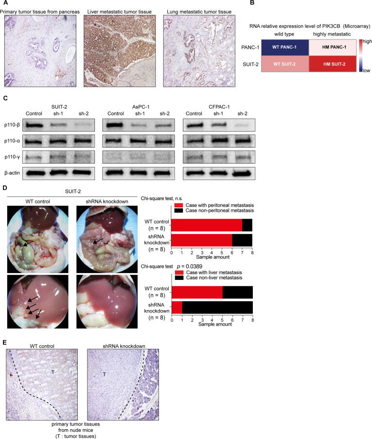
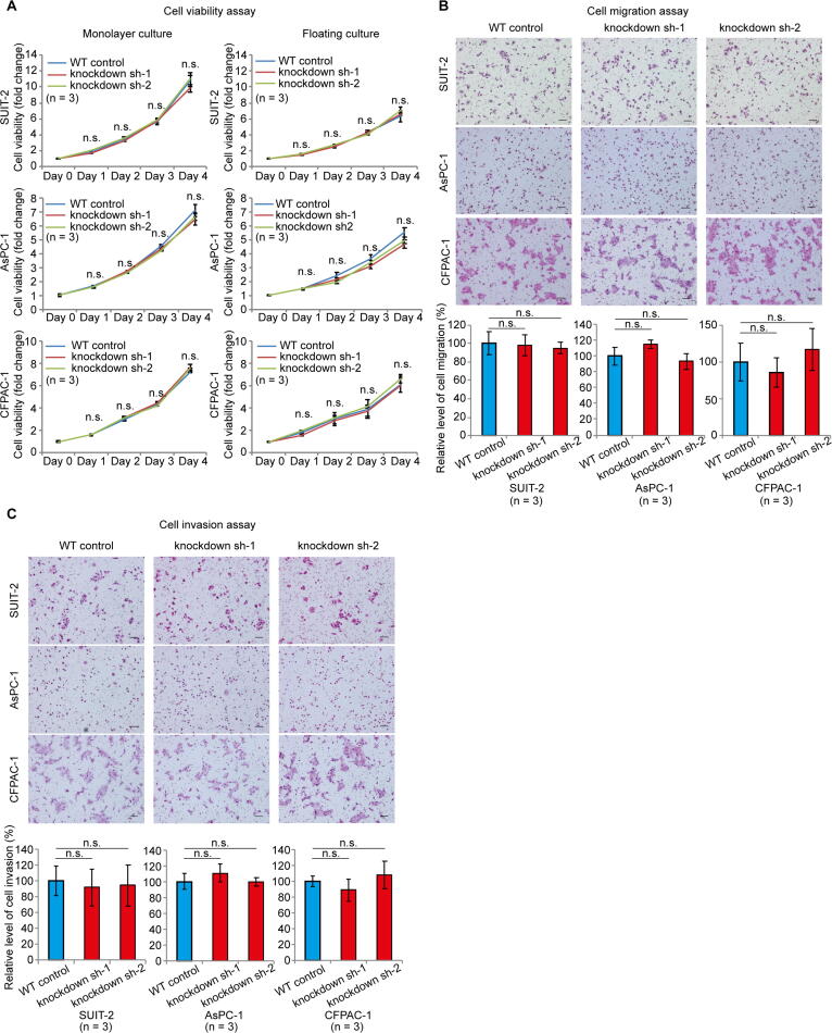
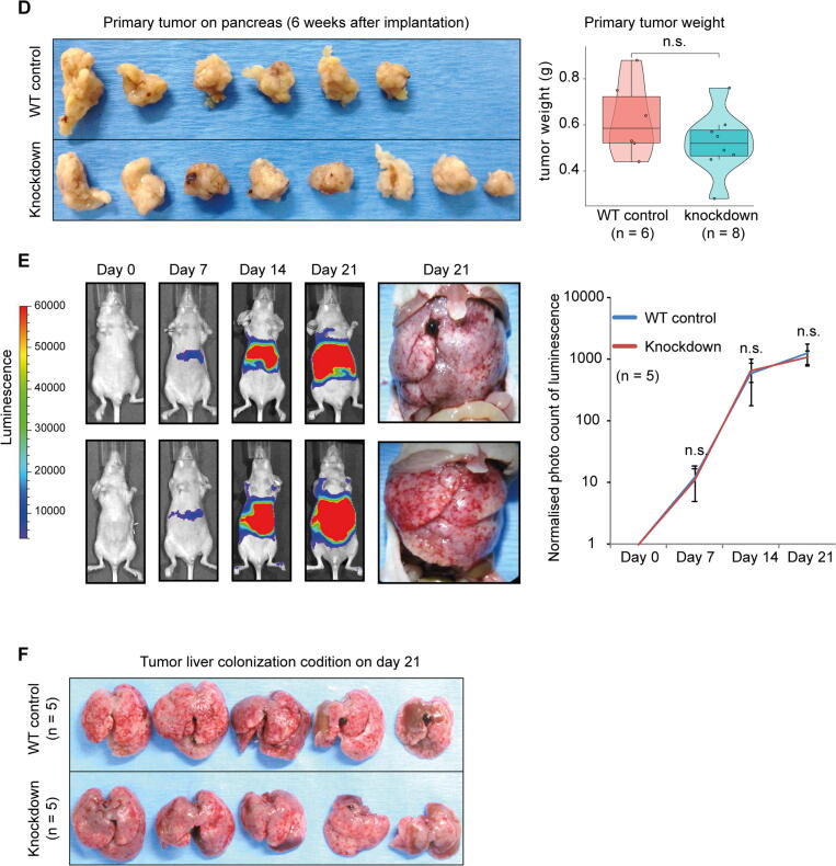
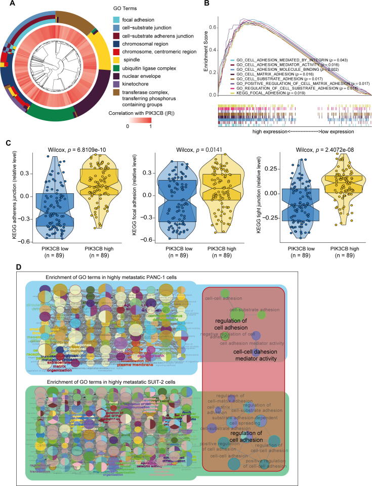
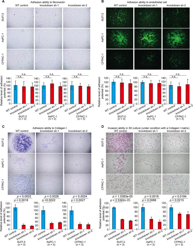
Results: We observed that PIK3CB was involved in PAAD progression. Notably, we identified that PIK3CB was involved in PAAD metastasis. Downregulation of PIK3CB significantly reduced PAAD metastatic potential in vivo. Furthermore, a series of bioinformatic analyses showed that PIK3CB was involved in cell adhesion in PAAD. Notably, PIK3CB depletion inhibited invasion potential specifically via suppressing cell adhesion to collagen I in PAAD cells.

Conclusion: Collectively, our findings indicate that PIK3CB is involved in PAAD metastasis through cell-matrix adhesion. We proposed that PIK3CB is a potential therapeutic target for PAAD therapy.

Keywords: Adhesion; Collagen I; Metastasis; PIK3CB; Pancreatic cancer.


Conflict of interest statement

The authors declare that they have no known competing financial interests or personal relationships that could have appeared to influence the work reported in this paper.

Figures

None
Graphical abstract
Fig. 1
Fig. 1
The expression pattern of PIK3CB in human PAAD. (A) PIK3CB RNA level in human PAAD was determined by GTEx-TCGA conjoint analysis and Arrayexpress analysis. (B) Assessment of the relationship between PIK3CB RNA expression and PAAD tumor histologic grade. (C) p110β IHC staining in pancreatic normal and tumor tissues. (D) Kaplan-Meier estimation for individuals with PAAD according to PIK3CB RNA expression. Datasets were download from ICGC database and KM-plotter database. (E) Kaplan-Meier estimation for individuals with PAAD according to p110β expression determined by IHC staining.
Fig. 2
Fig. 2
Assessment of relationship between PIK3CB expression and PAAD metastasis. (A) p110β IHC staining in tumor tissues from primary tumor and paired metastatic sites. (B) Identification of PIK3CB RNA expression in parental and highly metastatic PANC-1 and SUIT-2 cells determined by microarray. (C) Western blot was performed to examine p110β expression in parental and PIK3CB-depleted PAAD cells. (D) Murine model of PAAD peritoneal and hepatic metastasis was established by transplanting parental SUIT-2 or PIK3CB-depleted SUIT-2 cells into tail of pancreas in nude mice. Metastatic tumor tissues are indicated with black arrows. (E) IHC staining of p110β in parental SUIT-2 or PIK3CB-depleted SUIT-2 cells induced primary tumor tissues.
Fig. 3
Fig. 3
Comparison of cell viability, migration and invasion abilities between parental and PIK3CB-depleted PAAD cells. (A) Quantification of cell viability by continuous monitoring of parental and PIK3CB-deplated PAAD cells. (B) Assessment of cell migration ability in parental and PIK3CB-deplated PAAD cells. (C) Assessment of cell invasiveness in parental and PIK3CB-deplated PAAD cells. (D) Comparison of primary tumor growth (weight) between parental SUIT-2 induced tumors and PIK3CB-deplated SUIT-2 induced tumors. (E) Tumor hepatic colonization evaluation using the IVIS imaging system. (F) Tumor hepatic colonization on day 21 of IVIS examination.
Fig. 3
Fig. 3
Comparison of cell viability, migration and invasion abilities between parental and PIK3CB-depleted PAAD cells. (A) Quantification of cell viability by continuous monitoring of parental and PIK3CB-deplated PAAD cells. (B) Assessment of cell migration ability in parental and PIK3CB-deplated PAAD cells. (C) Assessment of cell invasiveness in parental and PIK3CB-deplated PAAD cells. (D) Comparison of primary tumor growth (weight) between parental SUIT-2 induced tumors and PIK3CB-deplated SUIT-2 induced tumors. (E) Tumor hepatic colonization evaluation using the IVIS imaging system. (F) Tumor hepatic colonization on day 21 of IVIS examination.
Fig. 4
Fig. 4
Functional annotation of PIK3CB in PAAD. (A) PIK3CB-related GO terms. Spearman’s correlation analysis was used to screen PIK3CB correlated genes according to the criteria that |R| > 0.4. Expression dataset from TCGA was used for the analysis. We carried out GO analysis to explore the biological function of PIK3CB using its correlated genes. (B) Enrichment blot from GSEA. We conducted GSEA between low and high PIK3CB expression tissues. The significantly changed cell adhesion related GO terms were selected and showed. The dataset from TCGA database was used for GSEA analysis. (C) Comparison of cell adhesion related pathways using GSVA. We conducted GSVA between low and high PIK3CB expression tissues. The significantly changed cell adhesion pathways (KEGG) were selected and showed. The dataset from TCGA database was used for GSVA analysis. (D) GO analysis of differential expression RNAs between parental and highly metastatic PAAD cells. Differentially expressed genes in highly metastatic PANC-1 and SUIT-2 cells were identified as fold change cutoff > 1, p < 0.05. Venn diagram representation showed that the differentially expressed genes are enriched in the cell adhesion related GO terms.
Fig. 5
Fig. 5
Cell adhesion evaluation in parental and PIK3CB-deplated PAAD cells. (A) Analysis of cell adhesion capacity to fibronectin. (B) Analysis of cell adhesion capacity to endothelial cell. Green fluorescent protein was expressed as a marker in PAAD cells. (C) Analysis of cell adhesion capacity to collagen I protein. (D) 3D invasion assay. The parental or PIK3CB-depleted PAAD cells were 3D cultured under condition with collagen I.
Fig. 6
Fig. 6
Predicted functional association of PIK3CB in cell adhesion. (A) Western blot analyses of total AKT protein and phosphorylation AKT protein in parental and PIK3CB-deplated PAAD cells. (B) Identification of possible interaction proteins of PIK3CB, The interaction datasets were downloaded from ComPPI database (http://comppi.linkgroup.hu/protein_search). R language was used for visualization of interaction proteins. The colours of lines indicated the localisation of interaction proteins. COL1A1 and COL1A2. Protein-protein interaction datasets were download from Compartmentalized Protein-Protein Interaction Database. (C) Protein-protein interaction analysis. The datasets were generated by string database (https://string-db.org/). Cytoscape software was used for data visualization.

Similar articles

Cited by

References

    1. Klein A.P., Wolpin B.M., Risch H.A., Stolzenberg-Solomon R.Z., Mocci E., Zhang M. Genome-wide meta-analysis identifies five new susceptibility loci for pancreatic cancer. Nat Commun. 2018;9. doi: 10.1038/s41467-018-02942-5. - DOI - PMC - PubMed
    1. Christenson E.S., Jaffee E., Azad N.S. Current and emerging therapies for patients with advanced pancreatic ductal adenocarcinoma: a bright future. Lancet Oncol. 2020;21:e135–e145. doi: 10.1016/S1470-2045(19)30795-8. - DOI - PMC - PubMed
    1. Rahib L., Smith B.D., Aizenberg R., Rosenzweig A.B., Fleshman J.M., Matrisian L.M. Projecting cancer incidence and deaths to 2030: the unexpected burden of thyroid, liver, and pancreas cancers in the united states. Cancer Res. 2014;74:2913–2921. doi: 10.1158/0008-5472.CAN-14-0155. - DOI - PubMed
    1. Winter J.M., Brennan M.F., Tang L.H., D’Angelica M.I., Dematteo R.P., Fong Y. Survival after resection of pancreatic adenocarcinoma: results from a single institution over three decades. Ann Surg Oncol. 2012;19:169–175. doi: 10.1245/s10434-011-1900-3. - DOI - PubMed
    1. Yachida S., Lacobuzio-Donahue C.A. The pathology and genetics of metastatic pancreatic cancer. Arch Pathol Lab Med. 2009;133:413–422. doi: 10.1043/1543-2165-133.3.413. - DOI - PubMed

Publication types