. 2021 Feb 9:33:215-225.
doi: 10.1016/j.jare.2021.01.016. eCollection 2021 Nov.

Longxuetongluo Capsule protects against cerebral ischemia/reperfusion injury through endoplasmic reticulum stress and MAPK-mediated mechanisms

Affiliations

Longxuetongluo Capsule protects against cerebral ischemia/reperfusion injury through endoplasmic reticulum stress and MAPK-mediated mechanisms

Bo Pan et al. J Adv Res. .

Abstract

Introduction: Longxuetongluo Capsule (LTC) is wildly applied to treat ischemic stroke in clinical practice in China. However, the pharmacological mechanism of LTC on ischemic stroke is still unstated.

Objective: Our research was designed to study the protective effect of LTC against cerebral ischemia-reperfusion (I/R) injury and reveal the underlying mechanism both in vivo and in vitro.

Methods: PC12 cells treated with glucose deprivation/reperfusion (OGD/R) were used to simulate in vitro ischemia/reperfusion (I/R) injury. The cell viability, apoptosis rate, and protein expressions of PC12 cells were evaluated. In vivo validation of the protective effect of LTC was carried out by middle cerebral artery occlusion (MCAO)/reperfusion treatment, and the underlying mechanism of its anti-apoptosis ability was further revealed by immunohistochemistry staining and Western blotting.

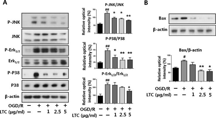
Results: In the current study, we observed that LTC effectively inhibited oxygen-glucose deprivation/reperfusion (OGD/R) induced apoptosis of PC12 cells through suppressing the cleavage of poly ADP-ribose polymerase (PARP), caspase-3, and caspase-9. Further investigation revealed that OGD/R insult remarkably triggered the endoplasmic reticulum stress responses (ER stress) to induce PC12 cell apoptosis. LTC treatment alleviated OGD/R induced ER stress by inhibiting the activation of protein kinase RNA (PKR)-like ER kinase (PERK)/eukaryotic translation initiation factor 2 (eIF2α) and inositol requiring enzyme 1 (IRE1)/tumor necrosis factor receptor-associated factor 2 (TRAF2) pathways. Additionally, LTC also restrained the OGD/R-induced PC12 cell apoptosis by reversing the activated mitogen-activated protein kinase (MAPK) through IRE1/TRAF2 pathway. Animal studies demonstrated LTC significantly restricted the infarct region induced by middle cerebral artery occlusion (MCAO)/reperfusion, the activation of ER stress and apoptosis of neuronal cells had also been suppressed by LTC in the penumbra region.

Conclusion: LTC protects the cerebral neuronal cell against ischemia/reperfusion injury through ER stress and MAPK-mediated mechanisms.

Keywords: Apoptosis; Endoplasmic reticulum stress; Longxuetongluo Capsule; Oxygen-glucose deprivation/reperfusion.


Conflict of interest statement

The authors declare that they have no known competing financial interests or personal relationships that could have appeared to influence the work reported in this paper.

Figures

None
Graphical abstract
Fig. 1
Fig. 1
LTC protected PC12 cells against oxygen-glucose deprivation/reperfusion (OGD/R)-induced cell apoptosis. (A) LTC improved the cell viability and reduced the LDH release of OGD/R-insulted PC12 cells. Cur: curcumin. (B) The effect of LTC alone on PC12 cell viability. (C) LTC suppressed the apoptosis rate of PC12 cell as measured by Annexin V staining and flow cytometry analysis. Q4: early apoptosis, Q2: late apoptosis. (D) Apoptotic PC12 cells were identified using AO/EB staining. Apoptotic cells were identified by double staining with acridine orange (AO) and ethidium bromide (EB). Cells that took up both dyes were classified as apoptotic (indicated by arrows in the EB and merged panels). Scale bar = 50 μm. All data are presented as Mean ± SEM, from independent experiments performed in triplicate, and statistical comparisons between the different groups were performed using one-way analysis of variance (ANOVA), followed by Student's t-test. ##P < 0.01, relative to untreated group; *P < 0.05, **P < 0.01, relative to OGD/R treated group.
Fig. 2
Fig. 2
LTC protected PC12 cells against OGD/R-mediated apoptosis by alleviating PERK- eIF2α mediated ER stress. (A) LTC reversed OGD/R-induced expression of cleaved PARP, caspase-3, and caspase-9. GAPDH was presented as the loading control. (B) OGD/R insult increased the expression of CHOP and the phosphorylation of PERK and eIF2αin PC12 cells at different time points. β-actin was presented as the loading control. (C) LTC markedly reversed the phosphorylation of PERK and eIF2α induced by OGD/R in a dose-dependent manner. (D) LTC reduced the expression of CHOP induced by OGD/R insult in PC12 cells. (E) LTC markedly reversed the phosphorylation of IRE1, Bip, and TRAF2 overexpression induced by OGD/R. Data are presented as Mean ± SEM, from independent experiments performed in triplicate ##P < 0.01, relative to untreated group; *P < 0.05, **P < 0.01, relative to OGD/R-treated group.
Fig. 3
Fig. 3
LTC down-regulated the activation of MAPK signaling pathway and Bax expression in PC12 cells. (A) LTC reversed OGD/R-induced phosphorylation of JNK, Erk1/2, and P38 dose-dependently. (B) LTC markedly reduced the expression of Bax induced by OGD/R insult. Data are presented as Mean ± SEM, from independent experiments performed in triplicate. ##P < 0.01, relative to untreated group; *P < 0.05, **P < 0.01, relative to OGD/R-treated group.
Fig. 4
Fig. 4
LTC inhibited thapsigargin-induced activation of ER stress and phosphorylation of P38 in vitro. (A) LTC inhibited PERK, eIF2α, and IRE1 phosphorylation induced by thapsigargin (TG) in PC12 cells. (B) LTC suppressed TG-induced CHOP expression in PC12 cells. (C) LTC reversed the TG-induced phosphorylation of P38 in a dose-dependent manner. (D) LTC suppressed TG-induced PC12 apoptosis. Data are presented as Mean ± SEM, from independent experiments performed in triplicate. ##P < 0.01, relative to untreated group; *P < 0.05, **P < 0.01, relative to OGD/R treated group.
Fig. 5
Fig. 5
LTC protected cerebral I/R injury in vivo. (A) LTC remarkably reduced the infarct size in the coronal section of the rat brain. SD rats were subjected to MCAO (1.5 h) with the treatment of LTC (300 mg/kg) or nimodipine (10 mg/kg) for 5 days. Then, the coronal brain sections were cut and stained with TTC. The red regions indicated viable brain tissue, whereas non-stained pale regions indicated infarct brain tissue (n = 8). Scale bar = 10 mm. Nim: nimodipine. (B) LTC significantly reversed the neuronal structure changes, CHOP expression, and neuronal cell apoptosis in the coronal section of the rat brain caused by MCAO. The coronal brain sections were cut and stained with HE, TUNEL, or the antibodies of CHOP and NeuN in IHC assay. Black arrows indicate vacuolation and karyopyknosis of the neuronal cell in the representative photomicrographs of HE staining. Red arrows indicate apoptotic vascular cells in the representative photomicrographs of TUNEL staining. Yellow arrows show the positive staining in the representative photomicrographs of the CHOP IHC assay. Green arrows show the positive staining of the neuronal cell in the representative photomicrographs of the NeuN IHC assay. Scale bar = 50 μm. (C) LTC reversed the expressions of CHOP, cleaved caspase-3 expression, and P-P38 in rat brain caused by MCAO (n = 6). Data are presented as Mean ± SEM. ##P < 0.01, relative to sham group; *P < 0.05, **P < 0.01, relative to middle cerebral artery occlusion group.
Fig. 6
Fig. 6
Proposed mechanism of the protective effect of LTC against cerebral I/R injury in neuronal cells.

References

    1. Marciniec M., Sapko K., Kulczynski M., Popek-Marciniec S., Szczepanska-Szerej A., Rejdak K. Non-traumatic cervical artery dissection and ischemic stroke: a narrative review of recent research. Clin Neurol Neurosur. 2019;187 - PubMed
    1. Perez-de-Puig I., Miro-Mur F., Ferrer-Ferrer M., Gelpi E., Pedragosa J., Justicia C. Neutrophil recruitment to the brain in mouse and human ischemic stroke. ACTA Neuropathol. 2015;129:239–257. - PubMed
    1. Wei K., Wan L., Liu J., Zhang B., Li X., Zhang Y. Downregulation of Trb3 protects neurons against apoptosis induced by global cerebral ischemia and reperfusion Injury in rats. Neuroscience. 2017;360:118–127. - PubMed
    1. Shu Q., Fan H., Li S.J., Zhou D., Ma W., Zhao X.Y. Protective effects of Progranulin against focal cerebral ischemia-reperfusion injury in rats by suppressing endoplasmic reticulum stress and NF-kappa B activation in reactive astrocytes. J Cell Biochem. 2018;119:6584–6597. - PubMed
    1. Pluquet O., Pourtier A., Abbadie C. The unfolded protein response and cellular senescence. A review in the theme: cellular mechanisms of endoplasmic reticulum stress signaling in health and disease. Am J Physiol-Cell Ph. 2015;308:415–425. - PubMed

Publication types

MeSH terms

Substances