

Cross-sectional visual comparison of remineralization efficacy of various agents on early smooth surface caries of primary teeth with swept source optical coherence tomography
- PMID: 34603951
- PMCID: PMC8473773
- DOI: 10.1016/j.jobcr.2021.09.006
Cross-sectional visual comparison of remineralization efficacy of various agents on early smooth surface caries of primary teeth with swept source optical coherence tomography
Abstract
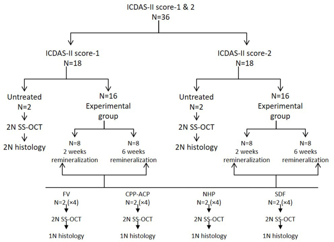
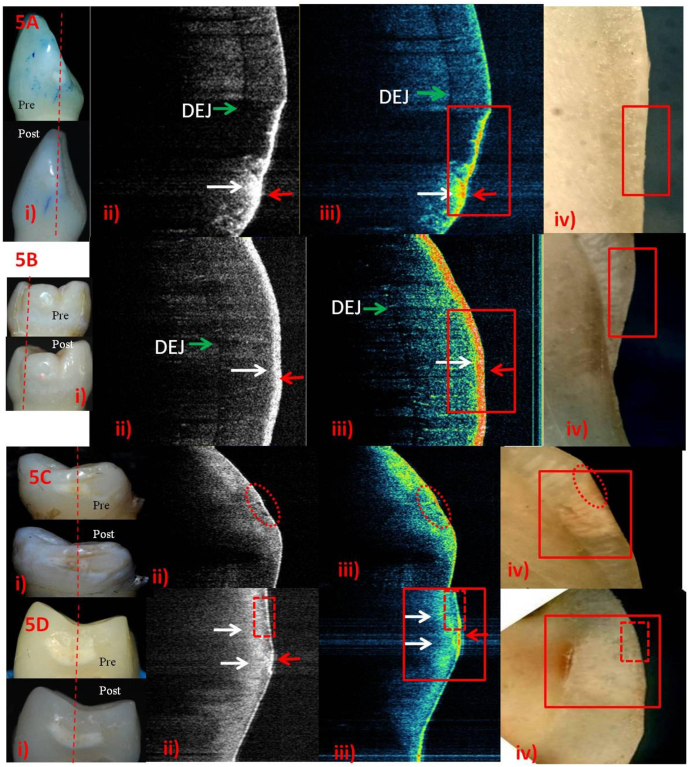
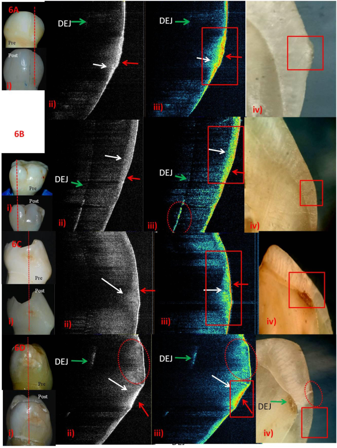
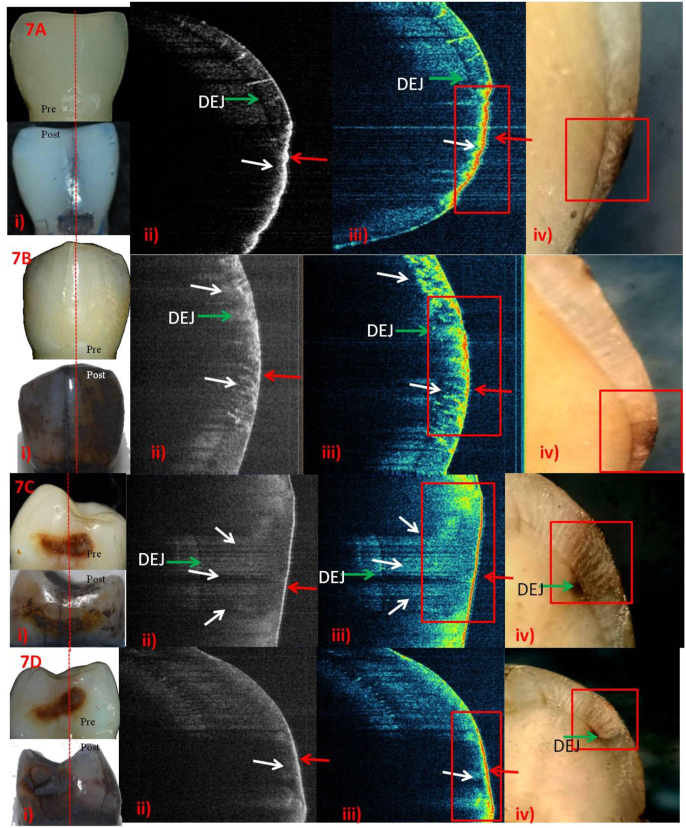
Purpose: Sweptsource optical coherence tomography (SS-OCT) permits cross-sectional observation of surface/subsurface characteristics of enamel including early carious lesions (ECL) or remineralization non-invasively.This study aimed to visually compare the cross-sectional remineralizing efficacy of various agents on ICDAS-II scores-1&2 by using SS-OCT and histology.
Methods: Baseline SS-OCT (grey-scale/false-colour) and histology was performed on the randomly selected two samples with scores-1&2. Four remineralizing agents [fluoride-varnish (FV), CPP-ACP, nanohydroxy-paste (NHP) and silver-diamine-fluoride (SDF)]were evaluated for 2-or 6-weeks post-remineralization using SS-OCT and histology.
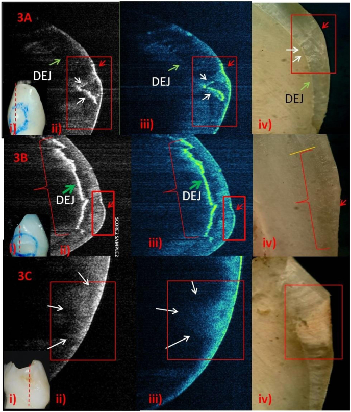
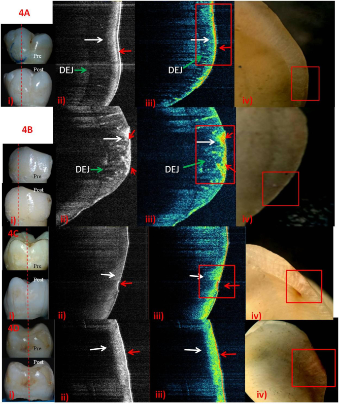
Results: Score-1&2 baseline SS-OCT images showed a linear-shaped demineralization with dentino-enamel junction (DEJ) visible; and bowl-shaped demineralization with DEJ invisible respectively. Remineralizing agents were assessed on the basis of their ability to remineralize the surface, subsurface and made visualize the DEJ in score-2. SS-OCT showed an outer growth layer in post-remineralization score-1, 2-weeks samples with FV and NHP. All the agents showed progressive subsurface remineralization in 6 weeks. Active lesions showed rapid uptake of minerals on surface. Subsurface mineralization in pigmented score-2 matched sound enamel with NHP and SDF. Surface remineralization was comparable in FV and SDF followed by NHP. SDF demonstrated deeper subsurface remineralization followed by NHP and CPP-ACP.
Conclusion: SS-OCT images correlated to histology. SS-OCT could monitor surface/subsurface in-situde/remineralization activity non-invasively.
Keywords: Early carious lesions; Enamel; ICDAS-II; Remineralization; SS-OCT; Smooth surface caries.
© 2021 Published by Elsevier B.V. on behalf of Craniofacial Research Foundation.
Figures
Similar articles
-
Comparative Evaluation of the Remineralizing Potential of Silver Diamine Fluoride, Casein Phosphopeptide-amorphous Calcium Phosphate, and Fluoride Varnish on the Enamel Surface of Primary and Permanent Teeth: An In Vitro Study.Int J Clin Pediatr Dent. 2023 Aug;16(Suppl 1):S91-S96. doi: 10.5005/jp-journals-10005-2622. Int J Clin Pediatr Dent. 2023. PMID: 37663209 Free PMC article.
-
Evaluation of Incipient Enamel Caries at Smooth Tooth Surfaces Using SS-OCT.Materials (Basel). 2022 Aug 28;15(17):5947. doi: 10.3390/ma15175947. Materials (Basel). 2022. PMID: 36079329 Free PMC article.
-
Remineralization potential of fluoride and amorphous calcium phosphate-casein phospho peptide on enamel lesions: An in vitro comparative evaluation.J Conserv Dent. 2010 Jan;13(1):42-6. doi: 10.4103/0972-0707.62634. J Conserv Dent. 2010. PMID: 20582219 Free PMC article.
-
Evaluation of dental caries, tooth crack, and age-related changes in tooth structure using optical coherence tomography.Jpn Dent Sci Rev. 2020 Nov;56(1):109-118. doi: 10.1016/j.jdsr.2020.08.001. Epub 2020 Oct 2. Jpn Dent Sci Rev. 2020. PMID: 33033549 Free PMC article. Review.
-
A Review of Casein Phosphopeptide-Amorphous Calcium Phosphate (CPP-ACP) and Enamel Remineralization.Compend Contin Educ Dent. 2016 Jan;37(1):36-9; quiz 40. Compend Contin Educ Dent. 2016. PMID: 26863219 Review.
Cited by
-
The Potential of Silver Diamine Fluoride in Non-Operative Management of Dental Caries in Primary Teeth: A Systematic Review.Medicina (Kaunas). 2024 Oct 23;60(11):1738. doi: 10.3390/medicina60111738. Medicina (Kaunas). 2024. PMID: 39596923 Free PMC article.
-
Improving occlusal caries detection using optical clearing agent for optical coherence tomography.Lasers Med Sci. 2025 Jun 12;40(1):275. doi: 10.1007/s10103-025-04532-6. Lasers Med Sci. 2025. PMID: 40500425
-
Remineralisation capability of silver diamine fluoride in artificial enamel lesions on smooth surfaces using quantitative light-induced fluorescence measurements in-vitro.Sci Rep. 2022 May 19;12(1):8498. doi: 10.1038/s41598-022-12498-6. Sci Rep. 2022. PMID: 35589795 Free PMC article.
References
-
- Vaswani S., Sharma D.S., Mishra S., Sharma S. Histologic validation of ICDAS‐II and polarization sensitive optical coherence tomography to detect smooth surface early carious lesions. Int J Pediatr Dent. 2019;29(2):193–202. - PubMed
-
- Ismail A.I., Sohn W., Tellez M. The International Caries Detection and Assessment System (ICDAS): an integrated system for measuring dental caries. Community Dent Oral Epidemiol. 2007;35:170–178. - PubMed
-
- Trevisan TC, Andrade MC, Presoto CD, Oliveira OB, Andrade MF, BortolattoJF.Hidden caries. A critical review. Sci J Dent2015;2:33-36.
-
- Bader J.D., Shugar D.A., Bonito A.J. Systematic reviews of selected dental caries diagnostic and management methods. J Dent Edu. 2001;65(10):961–968. - PubMed
LinkOut - more resources
Full Text Sources
