Review
. 2021 Sep 16:22:101600.
doi: 10.1016/j.jcot.2021.101600. eCollection 2021 Nov.

Imaging update on musculoskeletal infections

Affiliations
Review

Imaging update on musculoskeletal infections

Teck Yew Chin et al. J Clin Orthop Trauma. .

Abstract

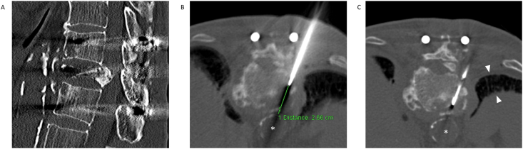
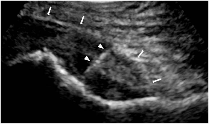
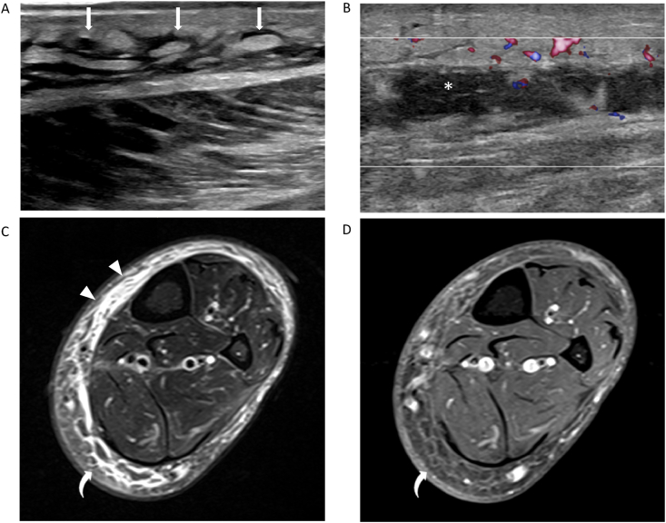
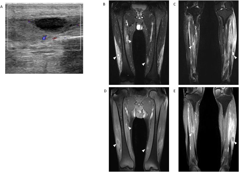
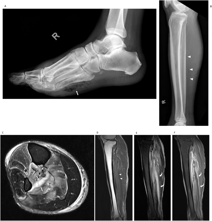
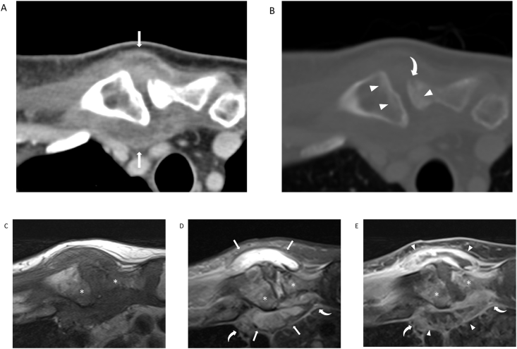
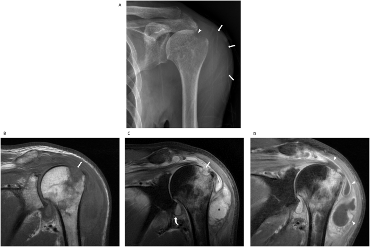
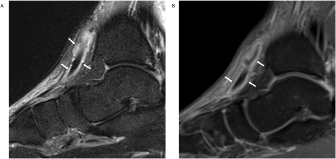
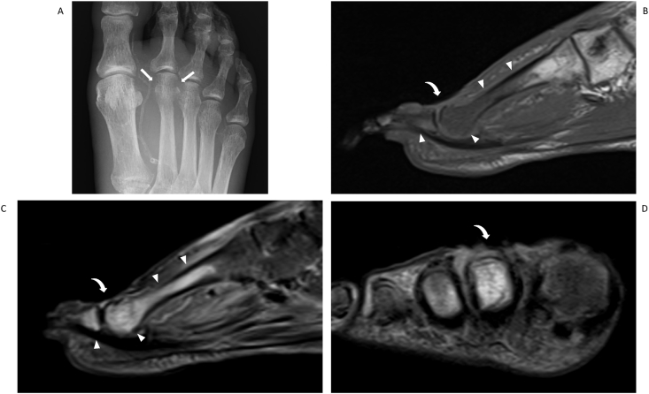
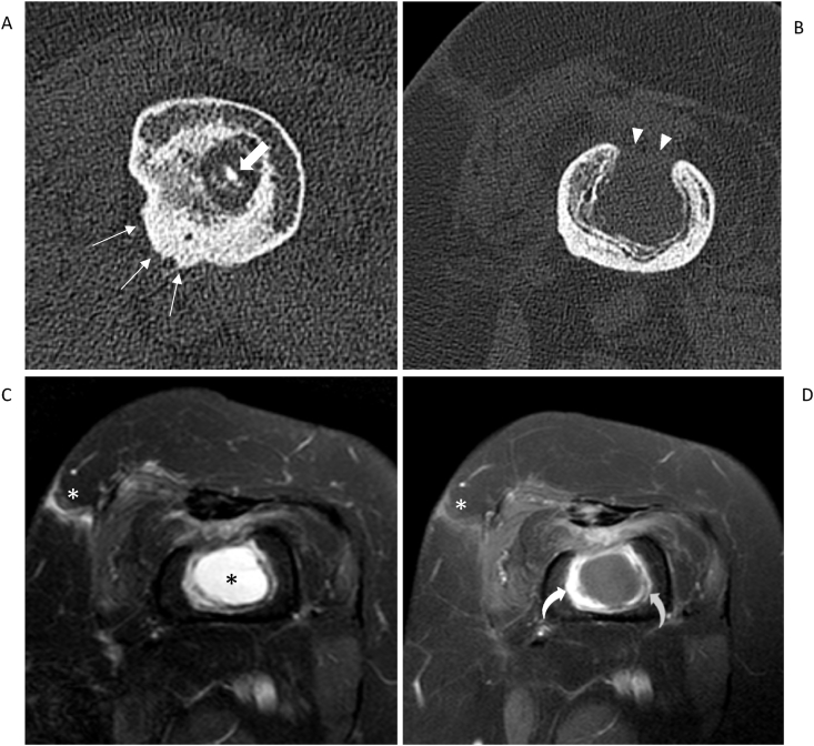
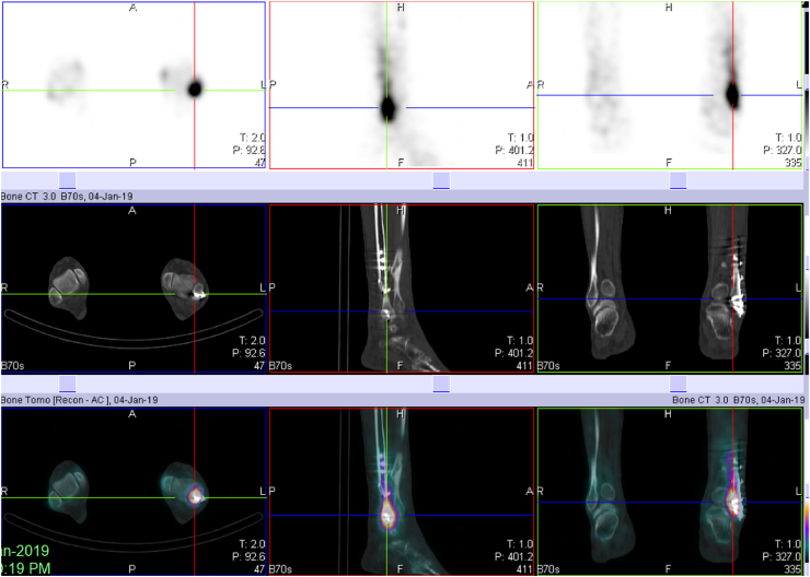
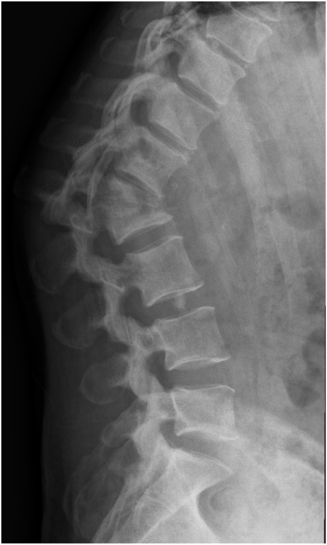
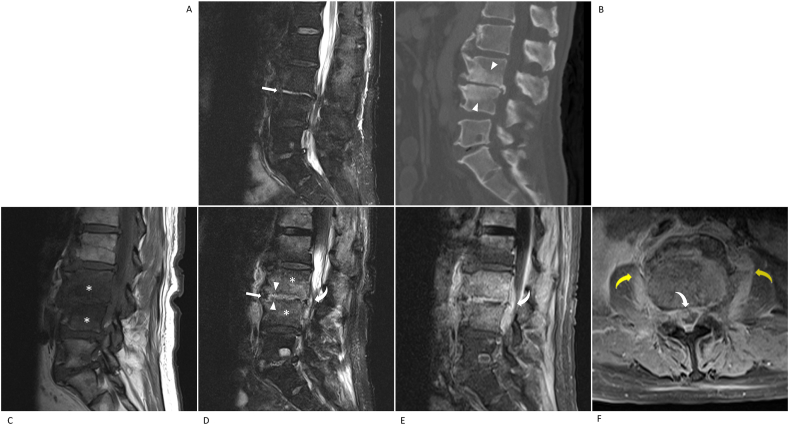
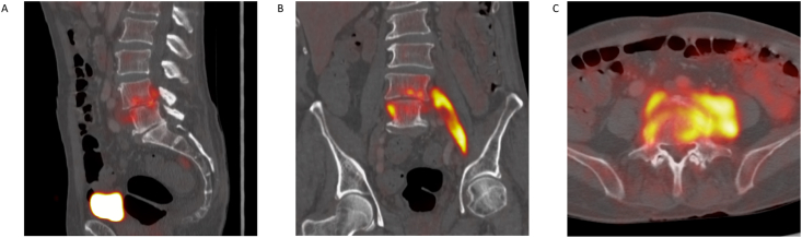
The clinical diagnosis of musculoskeletal infections can be challenging due to non-specific signs and symptoms on presentation. These infections include infectious myositis, necrotising fasciitis, septic arthritis, septic bursitis, suppurative tenosynovitis, osteomyelitis, spondylodiscitis and periprosthetic infections. Diagnostic imaging is routinely employed as part of the investigative pathway to characterise the underlying infectious disease pattern, allowing expedited and customised patient management plans to optimise outcomes. This article provides an update on the various imaging modalities comprising of radiography, computed tomography, ultrasonography, magnetic resonance imaging and radionuclide procedures, and incorporates representative images of key findings in the different forms of musculoskeletal infections.

Keywords: Musculoskeletal infection; Necrotising fasciitis; Osteomyelitis; Periprosthetic infections; Septic arthritis; Spondylodiscitis.


Figures

Fig. 1
Fig. 1
71-year-old woman who underwent posterior spinal stabilisation surgery for a T12 vertebral body burst fracture. Clinical progress was complicated by poor wound healing with increasing paraspinal soft tissue changes and clinical features of infection. (a) Sagittal CT image shows the vertebral burst fracture with retropulsion. (b,c) Axial CT-guided biopsy images using a costovertebral approach and avoiding critical structures like the aorta (∗) and lung (arrowheads). The tissue samples grew MRSA.
Fig. 2
Fig. 2
75-year-old woman presenting with left hip pain. US image shows an echogenic hip joint effusion (arrows) with needle placement for aspiration (arrowheads). MSSA was cultured from the joint aspirate.
Fig. 3
Fig. 3
66-year-old woman with diabetes mellitus presenting with erythema and right lower leg swelling. US images show (a) ‘cobblestone’ appearance of the subcutaneous fat (arrows) and (b) a linear pocket of hypoechoic fluid accumulation (asterisk) with mild surrounding hyperaemia. Axial fat-suppressed (c) T2-W and (D) contrast-enhanced T1-W MR images confirm the reticulated oedema pattern with areas of heterogeneous enhancement (curved arrows). Fluid accumulating along the peripheral layer of the deep intermuscular fascia (arrowheads) corresponds to the US image findings with no rim-enhancing abscess identified.
Fig. 4
Fig. 4
39-year-old man with undiagnosed and uncontrolled Type 2 diabetes mellitus. He presented with severe bilateral leg pain and mild left lower leg swelling. (a) Initial US image of the left lower leg shows multiple avascular, hypoechoic, and cystic-looking intramuscular lesions with a representative lesion as shown. Coronal fat-suppressed (b–c) T2-W MR images taken through the upper and lower leg, and corresponding (d–e) contrast-enhanced T1-W MR images show numerous intramuscular abscesses with surrounding muscle oedema (arrowheads). This is compatible with extensive pyomyositis. More than 15 intramuscular abscesses were peppered throughout the legs of this patient. The causative organism was MSSA.
Fig. 5
Fig. 5
A 50-year-old man, with diabetes mellitus and a right foot ulcer, presenting with septicaemia and increasing right lower leg pain. (a) Lateral radiograph of the foot shows subcutaneous emphysema over the plantar aspect of the foot from a gas-forming infection (arrow). (b) Lateral radiograph of the lower leg shows linear gas lucencies tracking along the deep intermuscular fascial planes (arrowheads) due to ascending necrotising fasciitis from the foot infection. (c) Axial fat-suppressed T2-W MR image shows thick deep intermuscular fluid between the tibialis posterior and flexor hallucis longus muscles (arrows) with extensive oedema of the surrounding muscles (asterisks) and surrounding cellulitis. Sagittal (d) T1-W, (e) fat-suppressed T2-W and (f) fat-suppressed contrast-enhanced T1-W MR images confirms rim enhancement of the fluid collection (curved arrows). Small punctate areas of signal loss correspond to the gas locules (arrowheads). The patient underwent surgical exploration and extensive tissue debridement, followed by negative pressure wound therapy.
Fig. 6
Fig. 6
78-year-old man with Group B Streptococcus septicaemia and right sternoclavicular joint pain. Axial contrast-enhanced CT images taken in (a) soft tissue and (b) bone window settings show an enhancing right sternoclavicular joint effusion (arrows) with subtle subarticular sclerosis (arrowheads) and a small osseous erosion at the sternal side (curved arrow). Axial (c) T1-W, (d) fat-suppressed T2-W and (e) fat-suppressed contrast-enhanced T1-W MR images show a joint effusion (arrows) with thick peripheral enhancement of the synovial capsule (arrowheads) and periarticular inflammation encroaching into the mediastinum (curved arrows). Subarticular bone marrow oedema and enhancement are also present (∗).
Fig. 7
Fig. 7
81-year-old woman presenting with left shoulder pain and swelling. (a) Frontal shoulder radiograph shows soft tissue swelling over the lateral aspect of the shoulder joint with loss of the normal fat planes (arrows). An osseous erosion is present at the greater tuberosity (arrowhead). Coronal (b) T1-W, (c) fat-suppressed T2-W and (d) fat-suppressed contrast-enhanced T1-W MR images show a large subacromial-subdeltoid bursal effusion (∗) with enhancement of the synovial bursal lining (arrowheads). The greater tuberosity osseous erosion (arrow) is appreciated with extensive surrounding bone marrow oedema. In this case, the bursal infection originated from the adjacent osteomyelitis of the greater tuberosity. A small non-specific glenohumeral effusion is also present (curved arrow). The causative organism was pan-sensitive S. aureus.
Fig. 8
Fig. 8
43-year-old woman with a ‘cat-scratch’ injury. Sagittal fat-suppressed (b) T2-W and (b) contrast-enhanced T1-W MR images show infectious tenosynovitis of the anterior tibialis tendon with tendon sheath effusion and enhancement (arrows).
Fig. 9
Fig. 9
75-year-old woman with diabetes mellitus and a right dorsal foot ulcer. (a) Frontal radiograph shows cortical erosions of the 2nd metatarsal head with adjacent osteopenia (arrows). Sagittal (b) T1-W, (c) fat-suppressed T2-W and (d) coronal fat-suppressed T2-W MR images show oedema seen as T2-hyperintensity with loss of the T1 intramedullary fat signal in the 2nd metatarsal and proximal phalanx (arrowheads). The overlying ulcer is appreciated (curved arrows).
Fig. 10
Fig. 10
63-year-old woman with a complex history of chronic osteomyelitis in the left femur. (a–b) Axial CT images show (a) a sequestrum (thick arrow) with an involucrum posteriorly (thin arrows) and (b) a large cloaca anteriorly (arrowheads). Axial fat-suppressed (c) T2-W and (d) contrast-enhanced T1-W MR images show a fluid collection (black asterisk) with enhancement of the surrounding granulation tissue (curved arrows). A partially visualized enhancing sinus tract is seen communicating to the skin surface (white asterisk).
Fig. 11
Fig. 11
SPECT/CT Tc99 m bone scan images show intense radiotracer uptake related to the lateral malleolus distal screws. Both aseptic and septic implant loosening can manifest in this manner.
Fig. 12
Fig. 12
52-year-old woman presenting with a 3-month history of worsening atraumatic lower back pain. Lateral radiograph shows a severe thoracolumbar junction kyphotic deformity from severe collapse and destruction of the L1 and L2 vertebral bodies, with loss of the intervertebral disc space. This is typical of a late presentation of spondylodiscitis.
Fig. 13
Fig. 13
74-year-old man with MSSA bacteriaemia and lower back pain. Initial sagittal (a) fat-suppressed T2-W MR and (c) CT images show isolated fluid in the L3/4 intervertebral disc (arrow) with chronic-looking degenerative endplate changes and sclerosis (arrowheads). 1 month follow-up sagittal (c) T1-W, (d) fat-suppressed T2-W and (e) fat-suppressed contrast-enhanced T1-W and (f) axial fat-suppressed contrast-enhanced T1-W MR images show that there is now established spondylodiscitis with vertebral body oedema (asterisks), progressive endplate irregularities (arrowheads), discal fluid (arrow), and epidural (white curved arrows) and paravertebral phlegmon (yellow curved arrows). There is pathological enhancement of the affected structures, particularly of the epidural and paravertebral soft tissue inflammation. The epidural phlegmon causes significant narrowing of the spinal canal.
Fig. 14
Fig. 14
Lumbar spondylodiscitis with psoas muscle involvement. (a) Sagittal, (b) coronal and (c) axial PET/CT images show increased uptake at the site of L4/5 spondylodiscitis with secondary left psoas muscle inflammation.

Similar articles

Cited by

References

    1. Chou E.H., Mann S., Hsu T.C. Incidence, trends, and outcomes of infection sites among hospitalizations of sepsis: a nationwide study. PloS One. 2020;15(1) doi: 10.1371/journal.pone.0227752. - DOI - PMC - PubMed
    1. Schwarz E.M., Parvizi J., Gehrke T. International consensus meeting on musculoskeletal infection: research priorities from the general assembly questions. J Orthop Res. 2018;37(5):997–1006. doi: 10.1002/jor.24293. May 2019. - DOI - PubMed
    1. Mathews C.J., Weston V.C., Jones A., Field M., Coakley G. Bacterial septic arthritis in adults. Lancet. 2010;375(9717):846–855. doi: 10.1016/s0140-6736(09)61595-6. Mar 6. - DOI - PubMed
    1. Raghavan M., Lazzeri E., Palestro C.J. Imaging of spondylodiscitis. Semin Nucl Med. 2018;48(2):131–147. doi: 10.1053/j.semnuclmed.2017.11.001. Mar. - DOI - PubMed
    1. Stevens D.L., Bryant A.E. Necrotizing soft-tissue infections. N Engl J Med. Dec 7 2017;377(23):2253–2265. doi: 10.1056/NEJMra1600673. - DOI - PubMed

LinkOut - more resources