Amide proton transfer (APT) imaging-based study on the correlation between brain pH and voltage-gated proton channels in piglets after hypoxic-ischemic brain injury
- PMID: 34603995
- PMCID: PMC8408782
- DOI: 10.21037/qims-21-250
Amide proton transfer (APT) imaging-based study on the correlation between brain pH and voltage-gated proton channels in piglets after hypoxic-ischemic brain injury
Abstract
Background: The normal regulation of brain pH is particularly critical for protein structure and enzymatic catalysis in the brain. This study aimed to investigate the regulation mechanism of brain pH after hypoxic-ischemic brain injury (HIBI) through the combination of amide proton transfer (APT) imaging, the analysis of brain pH levels, and the analysis of voltage-gated proton channel (Hv1) expression in piglets with HIBI.
Methods: A total of 59 healthy piglets (age range, 3-5 days after birth; body weight, 1-1.5 kg) were selected. Six piglets were excluded due to death, modeling failure, or motion artifacts, leaving a total of 10 animals in the control group and 43 animals in the HIBI model group. At different time points (0-2, 2-6, 6-12, 12-24, 24-48, and 48-72 hours) after HIBI, brain pH, Hv1 expression, and APT values were measured and analyzed. The statistical analysis of data was performed using the independent samples t-test, analysis of variance, and Spearman rank correlation analysis. A P value less than 0.05 indicated statistical significance.
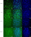
Results: As shown by the immunofluorescent staining results after HIBI, Hv1 protein expression in the basal ganglia reached a peak value at 0-2 hours, with a statistically significant difference between 0-2 hours and other time points (P<0.001). In piglets, the APT value reached a trough at 0-2 hours after HIBI, and subsequently, it gradually increased, and there was a significant difference between the control group and all HIBI model subgroups (P<0.001) except for the 2-6 hours subgroup (P=0.602). Brain pH decreased after HIBI and reached a trough at 0-2 hours, then gradually increased. Hv1 protein expression, pH, and APT values were all correlated (P<0.001).
Conclusions: After HIBI, values of brain pH, APT, and the expression of Hv1 changed over time and had a linear correlation. This suggests that there was a shift in brain hydrogen ions (H+) in the neural network and a change in brain pH after hypoxic-ischemic (HI) injury.
Keywords: Amide proton transfer (APT); brain; hypoxic-ischemic injury; pH; voltage-gated proton channels (Hv1).
2021 Quantitative Imaging in Medicine and Surgery. All rights reserved.
Conflict of interest statement
Conflicts of Interest: Both authors have completed the ICMJE uniform disclosure form (available at https://dx.doi.org/10.21037/qims-21-250). The authors have no conflicts of interest to declare.
Figures


Similar articles
-
Measurement of Lactate Content and Amide Proton Transfer Values in the Basal Ganglia of a Neonatal Piglet Hypoxic-Ischemic Brain Injury Model Using MRI.AJNR Am J Neuroradiol. 2017 Apr;38(4):827-834. doi: 10.3174/ajnr.A5066. Epub 2017 Feb 2. AJNR Am J Neuroradiol. 2017. PMID: 28154122 Free PMC article.
-
Deficiency in the voltage-gated proton channel Hv1 increases M2 polarization of microglia and attenuates brain damage from photothrombotic ischemic stroke.J Neurochem. 2016 Oct;139(1):96-105. doi: 10.1111/jnc.13751. Epub 2016 Sep 9. J Neurochem. 2016. PMID: 27470181 Free PMC article.
-
Voltage-gated proton channel HV1 in microglia.Neuroscientist. 2014 Dec;20(6):599-609. doi: 10.1177/1073858413519864. Epub 2014 Jan 23. Neuroscientist. 2014. PMID: 24463247 Review.
-
Early identification of neonatal mild hypoxic-ischemic encephalopathy by amide proton transfer magnetic resonance imaging: A pilot study.Eur J Radiol. 2019 Oct;119:108620. doi: 10.1016/j.ejrad.2019.07.021. Epub 2019 Jul 25. Eur J Radiol. 2019. PMID: 31422164
-
Functions and Mechanisms of the Voltage-Gated Proton Channel Hv1 in Brain and Spinal Cord Injury.Front Cell Neurosci. 2021 Apr 9;15:662971. doi: 10.3389/fncel.2021.662971. eCollection 2021. Front Cell Neurosci. 2021. PMID: 33897377 Free PMC article. Review.
Cited by
-
Amide proton transfer and apparent diffusion coefficient analysis reveal susceptibility of brain regions to neonatal hypoxic-ischemic encephalopathy.Heliyon. 2024 Sep 19;10(18):e38062. doi: 10.1016/j.heliyon.2024.e38062. eCollection 2024 Sep 30. Heliyon. 2024. PMID: 39347396 Free PMC article.
References
-
- Greco P, Nencini G, Piva I, Scioscia M, Volta CA, Spadaro S, Neri M, Bonaccorsi G, Greco F, Cocco I, Sorrentino F, D'Antonio F, Nappi L. Pathophysiology of hypoxic-ischemic encephalopathy: a review of the past and a view on the future. Acta Neurol Belg 2020;120:277-88. 10.1007/s13760-020-01308-3 - DOI - PubMed
-
- Khong PL, Tse C, Wong IY, Lam BC, Cheung PT, Goh WH, Kwong NS, Ooi GC. Diffusion-weighted imaging and proton magnetic resonance spectroscopy in perinatal hypoxic-ischemic encephalopathy: association with neuromotor outcome at 18 months of age. J Child Neurol 2004;19:872-81. 10.1177/08830738040190110501 - DOI - PubMed
LinkOut - more resources
Full Text Sources
Research Materials
