Uncommon presentation of craniospinal tuberculosis
- PMID: 34604017
- PMCID: PMC8472319
- DOI: 10.5339/qmj.2021.37
Uncommon presentation of craniospinal tuberculosis
Abstract
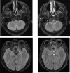
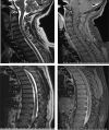
Tuberculosis (TB) is a bacterial infection with multisystem presentations. Involvement of the central nervous system (CNS) is considered the most lethal form among all types. In addition to possible fatality, CNS TB has serious neurological sequelae. These morbidity issues along with diagnostic challenges doubles the clinical burden. In recent years, there have been improvements in diagnostic sensitivity and specificity due to advances in technology. Herein, we report an atypical case of a patient with TB who presented to our department and discuss the flow of the diagnostic workup.
Keywords: craniospinal tuberculosis; uncommon presentation.
© 2021 Abdulrabu, Ebrahim, Warki, Alsotuhy, Anjum, licensee HBKU Press.
Figures


References
-
- Wilkinson RJ, Rohlwink U, Misra UK, van Crevel R, Mai NTH, Dooley KE, et al. Tuberculous meningitis. Nat Rev Neurol. 2017;13(10):581–598. - PubMed
-
- Thwaites GE, van Toorn R, Schoeman J. Tuberculous meningitis: more questions, still too few answers. Lancet Neurol. 2013;12(10):999–1010. - PubMed
-
- World Health Organization. Global tuberculosis report 2020 [Internet]. Geneva: World Health Organization; 2020. Available from: https://www.who.int/publications/i/item/9789240013131.
-
- Chin JH, Mateen FJ. Central nervous system tuberculosis: challenges and advances in diagnosis and treatment. Curr Infect Dis Rep. 2013;15(6):631–5. - PubMed
Publication types
LinkOut - more resources
Full Text Sources
