

Ceramide Regulates Anti-Tumor Mechanisms of Erianin in Androgen-Sensitive and Castration-Resistant Prostate Cancers
- PMID: 34604081
- PMCID: PMC8484793
- DOI: 10.3389/fonc.2021.738078
Ceramide Regulates Anti-Tumor Mechanisms of Erianin in Androgen-Sensitive and Castration-Resistant Prostate Cancers
Abstract
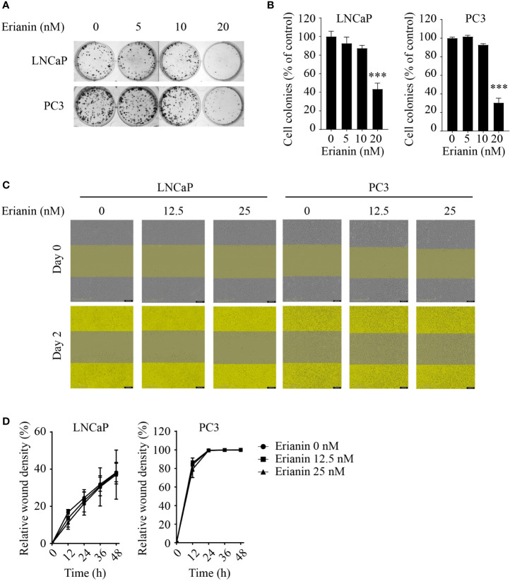
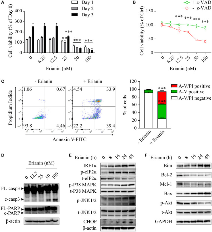
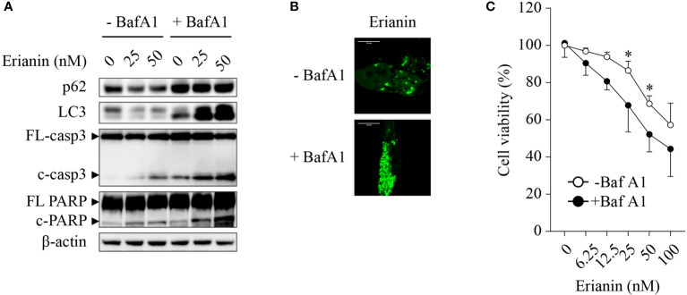
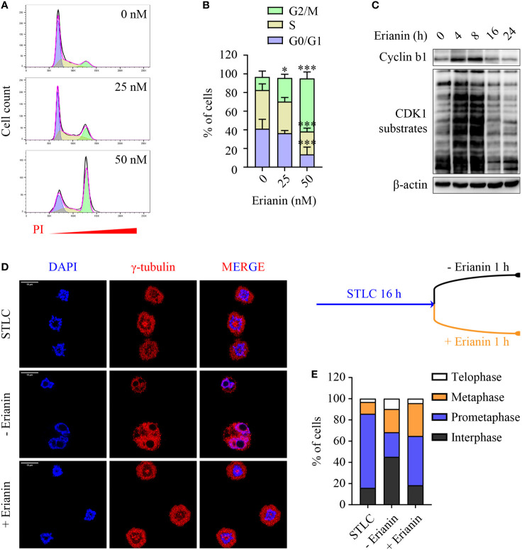
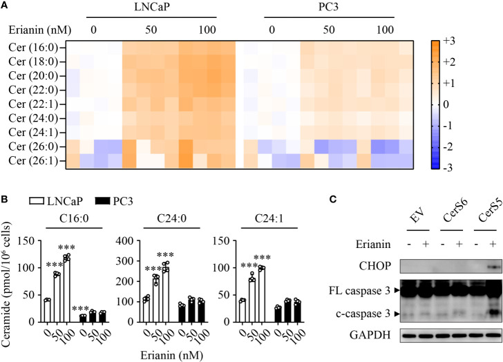
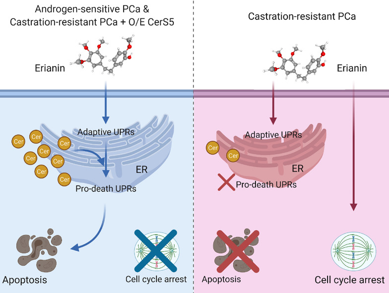
Prostate cancer is the second most prevalent malignancy worldwide. In the early stages, the development of prostate cancer is dependent on androgens. Over time with androgen deprivation therapy, 20% of prostate cancers progress to a castration-resistant form. Novel treatments for prostate cancers are still urgently needed. Erianin is a plant-derived bibenzyl compound. We report herein that erianin exhibits anti-tumor effects in androgen-sensitive and castration-resistant prostate cancer cells through different mechanisms. Erianin induces endoplasmic reticulum stress-associated apoptosis in androgen-sensitive prostate cancer cells. It also triggers pro-survival autophagic responses, as inhibition of autophagy predisposes to apoptosis. In contrast, erianin fails to induce apoptosis in castration-resistant prostate cancer cells. Instead, it results in cell cycle arrest at the M phase. Mechanistically, C16 ceramide dictates differential responses of androgen-sensitive and castration-resistant prostate cancer cells to erianin. Erianin elevates C16 ceramide level in androgen-sensitive but not castration-resistant prostate cancer cells. Overexpression of ceramide synthase 5 that specifically produces C16 ceramide enables erianin to induce apoptosis in castration-resistant prostate cancer cells. Our study provides both experimental evidence and mechanistic data showing that erianin is a potential treatment option for prostate cancers.
Keywords: apoptosis; autophagy; cell cycle arrest; endoplasmic reticulum stress; sphingolipid.
Copyright © 2021 Trapika, Liu, Chung, Lai, Xie, Zhao, Cui, Chen, Tran, Wang, Zhang, Don, Li, Hanrahan and Qi.
Conflict of interest statement
The authors declare that the research was conducted in the absence of any commercial or financial relationships that could be construed as a potential conflict of interest.
Figures

Similar articles
-
Nelfinavir inhibits regulated intramembrane proteolysis of sterol regulatory element binding protein-1 and activating transcription factor 6 in castration-resistant prostate cancer.FEBS J. 2012 Jul;279(13):2399-411. doi: 10.1111/j.1742-4658.2012.08619.x. Epub 2012 May 21. FEBS J. 2012. PMID: 22540830
-
C16 ceramide accumulates following androgen ablation in LNCaP prostate cancer cells.Prostate. 2003 Sep 15;57(1):66-79. doi: 10.1002/pros.10275. Prostate. 2003. PMID: 12886525
-
[COST ANALYSIS OF ANDROGEN DEPRIVATION THERAPY AND DRUGS FOR CASTRATION-RESISTANT PROSTATE CANCER].Nihon Hinyokika Gakkai Zasshi. 2021;112(2):53-57. doi: 10.5980/jpnjurol.112.53. Nihon Hinyokika Gakkai Zasshi. 2021. PMID: 35444081 Japanese.
-
Timing of androgen deprivation monotherapy and combined treatments in castration-sensitive and castration-resistant prostate cancer: a narrative review.World J Urol. 2020 Mar;38(3):601-611. doi: 10.1007/s00345-019-02704-y. Epub 2019 Mar 4. World J Urol. 2020. PMID: 30830274 Review.
-
Experimental evidence of persistent androgen-receptor-dependency in castration-resistant prostate cancer.Int J Mol Sci. 2013 Jul 26;14(8):15615-35. doi: 10.3390/ijms140815615. Int J Mol Sci. 2013. PMID: 23896594 Free PMC article. Review.
Cited by
-
Erianin Impedes the Proliferation and Metastatic Migration Through Suppression of STAT-3 Phosphorylation in Human Esophageal Cancer Cells.Appl Biochem Biotechnol. 2024 Sep;196(9):5859-5874. doi: 10.1007/s12010-023-04829-8. Epub 2024 Jan 2. Appl Biochem Biotechnol. 2024. PMID: 38165593
-
Ceramide Transfer Protein (CERT): An Overlooked Molecular Player in Cancer.Int J Mol Sci. 2021 Dec 7;22(24):13184. doi: 10.3390/ijms222413184. Int J Mol Sci. 2021. PMID: 34947980 Free PMC article. Review.
-
The Plant Derived 3-3'-Diindolylmethane (DIM) Behaves as CB2 Receptor Agonist in Prostate Cancer Cellular Models.Int J Mol Sci. 2023 Feb 11;24(4):3620. doi: 10.3390/ijms24043620. Int J Mol Sci. 2023. PMID: 36835033 Free PMC article.
-
The roles of ERIANIN in tumor and innate immunity and its' perspectives in immunotherapy.Front Immunol. 2023 Apr 28;14:1170754. doi: 10.3389/fimmu.2023.1170754. eCollection 2023. Front Immunol. 2023. PMID: 37187758 Free PMC article. Review.
-
Research progress on the regulatory and pharmacological mechanism of chemical components of Dendrobium.Heliyon. 2024 Sep 7;10(18):e37541. doi: 10.1016/j.heliyon.2024.e37541. eCollection 2024 Sep 30. Heliyon. 2024. PMID: 39328574 Free PMC article. Review.
References
LinkOut - more resources
Full Text Sources