Circular RNA 0006349 Augments Glycolysis and Malignance of Non-small Cell Lung Cancer Cells Through the microRNA-98/MKP1 Axis
- PMID: 34604211
- PMCID: PMC8484757
- DOI: 10.3389/fcell.2021.690307
Circular RNA 0006349 Augments Glycolysis and Malignance of Non-small Cell Lung Cancer Cells Through the microRNA-98/MKP1 Axis
Abstract
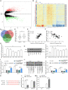
Background: The involvement of dysregulated circular RNAs (circRNAs) in human diseases has been increasingly recognized. In this study, we focused on the function of a newly screened circRNA, circ_0006349, in the progression of non-small-cell lung cancer (NSCLC) and the molecules of action. Methods: The NSCLC circRNA dataset GSE101684, microRNA (miRNA) dataset GSE29250, and mRNA dataset GSE51852 obtained from the GEO database were used to identify the differentially expressed genes in NSCLC samples. Tumor and normal tissues were collected from 59 patients with NSCLC. The expression of circ_0006349, miR-98, and MAP kinase phosphatase 1 (MKP1) in collected tissue samples and in acquired cells was determined. The binding relationships between miR-98 and circ_0006349/MKP1 were predicted and validated. Altered expression of circ_0006349, miR-98, and MKP1 was introduced in NSCLC cells to examine their roles in cell growth, apoptosis, and glycolysis. Results: Circ_0006349 and MKP1 were upregulated, and miR-98 was poorly expressed in the collected tumor tissues and the acquired NSCLC cell lines. Circ_0006349 was identified as a sponge for miR-98 to elevate MKP1 expression. Silencing of circ_0006349 suppressed proliferation and increased apoptosis of Calu-3 and H1299 cells, and it reduced glycolysis, glucose uptake, and the production of lactate in cells. Upon circ_0006349 knockdown, further downregulation of miR-98 or upregulation of MKP1 restored the malignant behaviors of cells. Conclusion: This research demonstrated that circ_0006349 derepressed MKP1 expression by absorbing miR-98, which augmented the proliferation and glycolysis of NSCLC cells and promoted cancer development.
Keywords: Circ_0006349; MKP1; NSCLC; glycolysis; miR-98; proliferation.
Copyright © 2021 Qin, Lu, Yuan, Zhao, Zhou, Fan, Yin, Yu and Bian.
Conflict of interest statement
The authors declare that the research was conducted in the absence of any commercial or financial relationships that could be construed as a potential conflict of interest.
Figures


Similar articles
-
Depletion of circ-BIRC6, a circular RNA, suppresses non-small cell lung cancer progression by targeting miR-4491.Biosci Trends. 2021 Jan 23;14(6):399-407. doi: 10.5582/bst.2020.03310. Epub 2020 Nov 10. Biosci Trends. 2021. PMID: 33177288
-
CircRNA circ_0006677 Inhibits the Progression and Glycolysis in Non-Small-Cell Lung Cancer by Sponging miR-578 and Regulating SOCS2 Expression.Front Pharmacol. 2021 May 13;12:657053. doi: 10.3389/fphar.2021.657053. eCollection 2021. Front Pharmacol. 2021. PMID: 34054537 Free PMC article.
-
Circ_0014130 Participates in the Proliferation and Apoptosis of Nonsmall Cell Lung Cancer Cells via the miR-142-5p/IGF-1 Axis.Cancer Biother Radiopharm. 2020 Apr;35(3):233-240. doi: 10.1089/cbr.2019.2965. Epub 2020 Jan 9. Cancer Biother Radiopharm. 2020. PMID: 31916848
-
Suppression of Circular RNA Hsa_circ_0109320 Attenuates Non-Small Cell Lung Cancer Progression via MiR-595/E2F7 Axis.Med Sci Monit. 2020 Jun 8;26:e921200. doi: 10.12659/MSM.921200. Med Sci Monit. 2020. PMID: 32508344 Free PMC article.
-
circ_0007385 served as competing endogenous RNA for miR-519d-3p to suppress malignant behaviors and cisplatin resistance of non-small cell lung cancer cells.Thorac Cancer. 2020 Aug;11(8):2196-2208. doi: 10.1111/1759-7714.13527. Epub 2020 Jun 29. Thorac Cancer. 2020. PMID: 32602212 Free PMC article.
Cited by
-
Knockdown of Glycolysis-Related LINC01070 Inhibits the Progression of Breast Cancer.Cureus. 2024 May 11;16(5):e60093. doi: 10.7759/cureus.60093. eCollection 2024 May. Cureus. 2024. PMID: 38860098 Free PMC article.
-
Research Progress of circRNAs in Glioblastoma.Front Cell Dev Biol. 2021 Nov 22;9:791892. doi: 10.3389/fcell.2021.791892. eCollection 2021. Front Cell Dev Biol. 2021. PMID: 34881248 Free PMC article. Review.
-
circCAPRIN1 interacts with STAT2 to promote tumor progression and lipid synthesis via upregulating ACC1 expression in colorectal cancer.Cancer Commun (Lond). 2023 Jan;43(1):100-122. doi: 10.1002/cac2.12380. Epub 2022 Nov 3. Cancer Commun (Lond). 2023. PMID: 36328987 Free PMC article.
-
The Metabolism Reprogramming of microRNA Let-7-Mediated Glycolysis Contributes to Autophagy and Tumor Progression.Int J Mol Sci. 2021 Dec 22;23(1):113. doi: 10.3390/ijms23010113. Int J Mol Sci. 2021. PMID: 35008539 Free PMC article. Review.
-
CircPKM2 aggravates the progression of non-small cell lung cancer by regulating MTDH via miR-1298-5p.Thorac Cancer. 2023 Oct;14(30):3020-3031. doi: 10.1111/1759-7714.15092. Epub 2023 Sep 7. Thorac Cancer. 2023. PMID: 37675591 Free PMC article.
References
LinkOut - more resources
Full Text Sources
Miscellaneous

