

Primordial Germ Cell Specification in Vertebrate Embryos: Phylogenetic Distribution and Conserved Molecular Features of Preformation and Induction
- PMID: 34604230
- PMCID: PMC8481613
- DOI: 10.3389/fcell.2021.730332
Primordial Germ Cell Specification in Vertebrate Embryos: Phylogenetic Distribution and Conserved Molecular Features of Preformation and Induction
Abstract
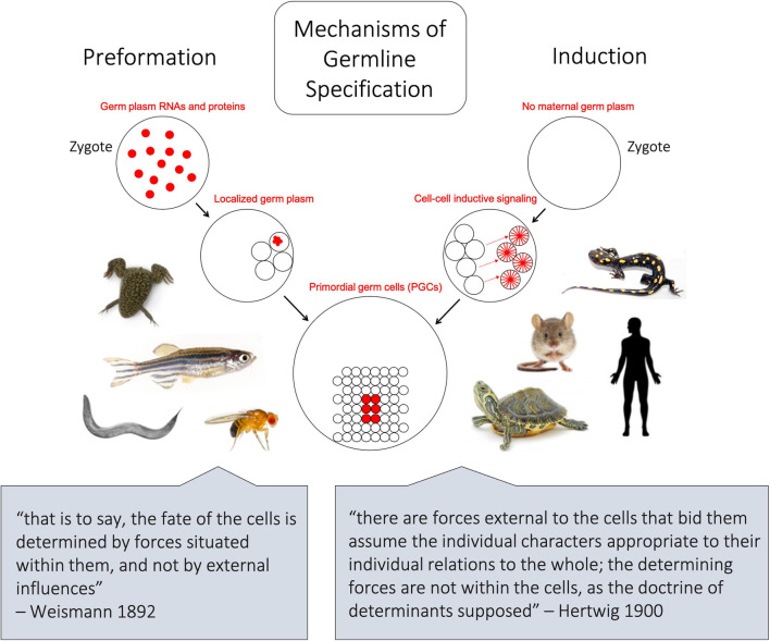
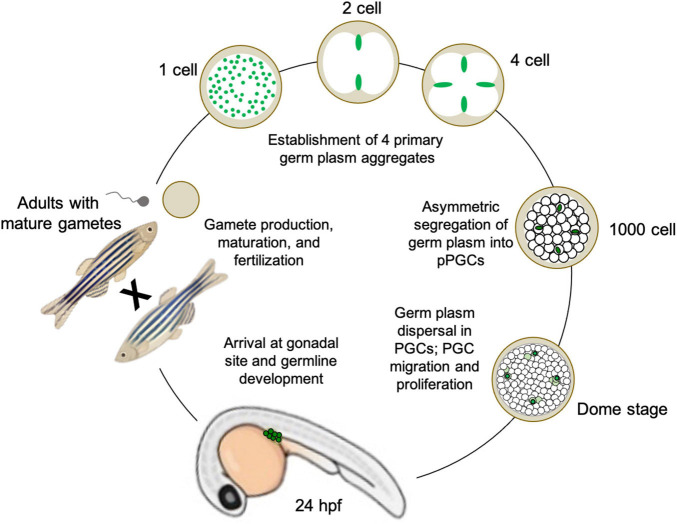
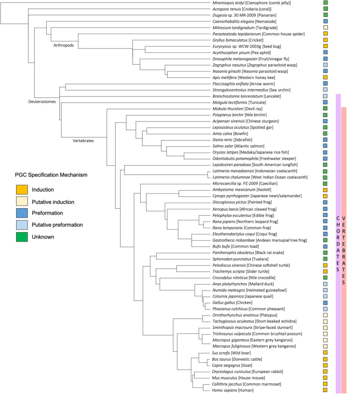
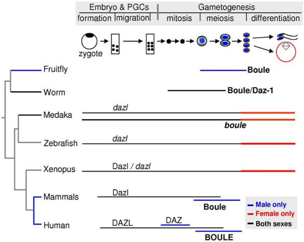
The differentiation of primordial germ cells (PGCs) occurs during early embryonic development and is critical for the survival and fitness of sexually reproducing species. Here, we review the two main mechanisms of PGC specification, induction, and preformation, in the context of four model vertebrate species: mouse, axolotl, Xenopus frogs, and zebrafish. We additionally discuss some notable molecular characteristics shared across PGC specification pathways, including the shared expression of products from three conserved germline gene families, DAZ (Deleted in Azoospermia) genes, nanos-related genes, and DEAD-box RNA helicases. Then, we summarize the current state of knowledge of the distribution of germ cell determination systems across kingdom Animalia, with particular attention to vertebrate species, but include several categories of invertebrates - ranging from the "proto-vertebrate" cephalochordates to arthropods, cnidarians, and ctenophores. We also briefly highlight ongoing investigations and potential lines of inquiry that aim to understand the evolutionary relationships between these modes of specification.
Keywords: embryonic development; germ cells; germline specification; induction; preformation; primordial germ cells (PGCs).
Copyright © 2021 Hansen and Pelegri.
Conflict of interest statement
The authors declare that the research was conducted in the absence of any commercial or financial relationships that could be construed as a potential conflict of interest.
Figures


Similar articles
-
Stochastic specification of primordial germ cells from mesoderm precursors in axolotl embryos.Development. 2014 Jun;141(12):2429-40. doi: 10.1242/dev.105346. Development. 2014. PMID: 24917499 Free PMC article.
-
Expression of germline markers in three species of amphioxus supports a preformation mechanism of germ cell development in cephalochordates.Evodevo. 2013 Jun 18;4(1):17. doi: 10.1186/2041-9139-4-17. Evodevo. 2013. PMID: 23777831 Free PMC article.
-
Complexities in Bombyx germ cell formation process revealed by Bm-nosO (a Bombyx homolog of nanos) knockout.Dev Biol. 2019 Jan 1;445(1):29-36. doi: 10.1016/j.ydbio.2018.10.012. Epub 2018 Oct 24. Dev Biol. 2019. PMID: 30367845
-
Germline development in amniotes: A paradigm shift in primordial germ cell specification.Bioessays. 2016 Aug;38(8):791-800. doi: 10.1002/bies.201600025. Epub 2016 Jun 6. Bioessays. 2016. PMID: 27273724 Free PMC article. Review.
-
Mechanisms of Vertebrate Germ Cell Determination.Adv Exp Med Biol. 2017;953:383-440. doi: 10.1007/978-3-319-46095-6_8. Adv Exp Med Biol. 2017. PMID: 27975276 Review.
Cited by
-
In vitro oogenesis from murine premeiotic germ cells using a new three-dimensional culture system.Cell Death Discov. 2023 Jul 31;9(1):276. doi: 10.1038/s41420-023-01577-w. Cell Death Discov. 2023. PMID: 37518361 Free PMC article.
-
In Vitro models of leukemia development: the role of very small leukemic stem-like cells in the cellular transformation cascade.Front Cell Dev Biol. 2025 Jan 3;12:1463807. doi: 10.3389/fcell.2024.1463807. eCollection 2024. Front Cell Dev Biol. 2025. PMID: 39830209 Free PMC article. Review.
-
DMRT1 regulates human germline commitment.Nat Cell Biol. 2023 Oct;25(10):1439-1452. doi: 10.1038/s41556-023-01224-7. Epub 2023 Sep 14. Nat Cell Biol. 2023. PMID: 37709822 Free PMC article.
-
Novel cell states arise in embryonic cells devoid of key reprogramming factors.bioRxiv [Preprint]. 2024 May 15:2024.05.13.593729. doi: 10.1101/2024.05.13.593729. bioRxiv. 2024. PMID: 38798464 Free PMC article. Preprint.
-
Essential functions of RNA helicase Vasa in maintaining germline stem cells and piRNA-guided Stellate silencing in Drosophila spermatogenesis.Front Cell Dev Biol. 2024 Aug 9;12:1450227. doi: 10.3389/fcell.2024.1450227. eCollection 2024. Front Cell Dev Biol. 2024. PMID: 39184915 Free PMC article.
References
-
- Akam M. (1987). The molecular basis for metameric pattern in the Drosophila embryo. Development (Camb. Engl.) 101 1–22. - PubMed
-
- Al-Mukhtar K., Webb A. (1971). An Ultrastructural study of primordial germ cells, oogonia and early oocytes in Xenopus Laevis. J. Embryol. Exp. Morphol. 26 195–217. - PubMed
Publication types
Grants and funding
LinkOut - more resources
Full Text Sources