. 2021 Sep 17:9:736603.
doi: 10.3389/fcell.2021.736603. eCollection 2021.

IL-13 Alleviates Cardiomyocyte Apoptosis by Improving Fatty Acid Oxidation in Mitochondria

Affiliations

IL-13 Alleviates Cardiomyocyte Apoptosis by Improving Fatty Acid Oxidation in Mitochondria

Xiaoyu Guo et al. Front Cell Dev Biol. .

Abstract

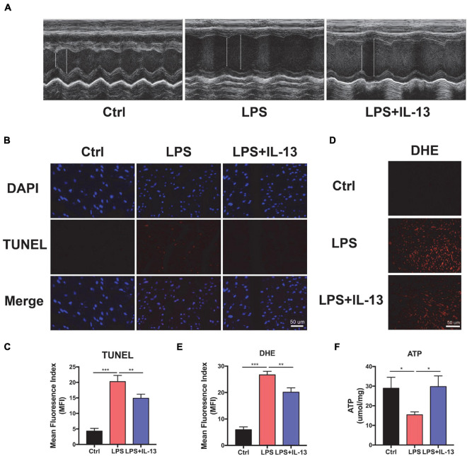
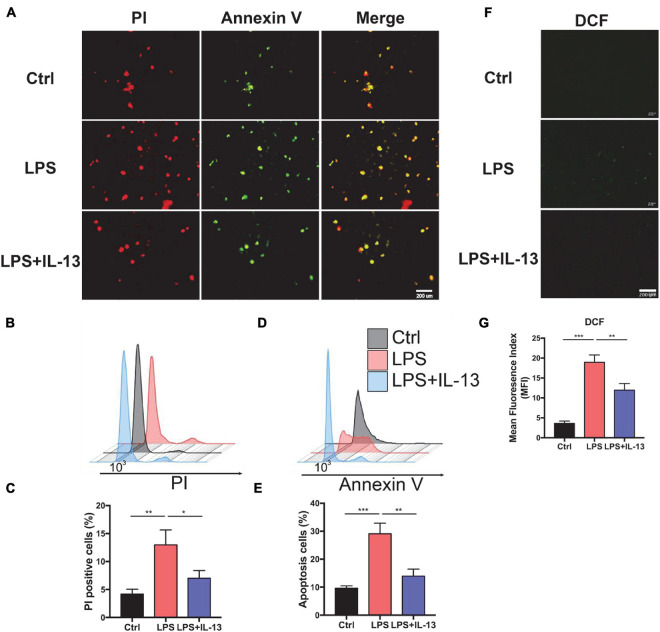
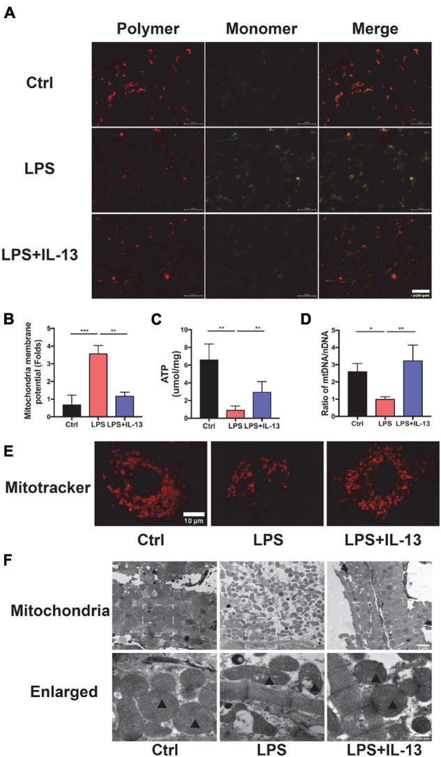
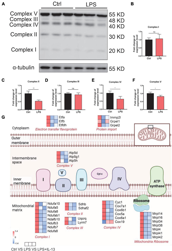
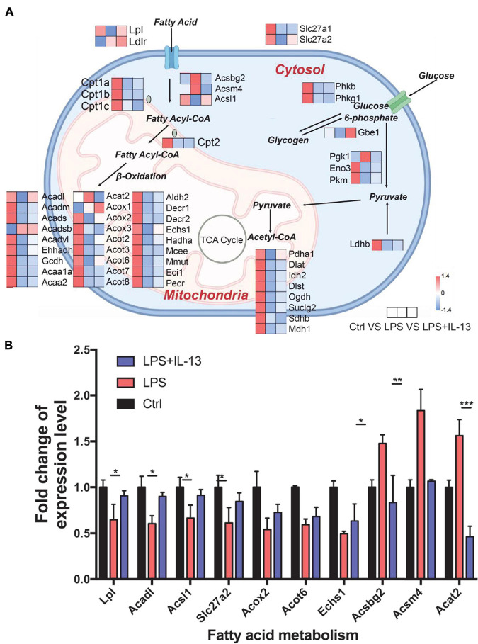
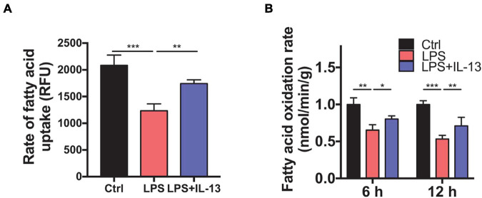
Sepsis-induced cardiac injury (SIC) is one of the most common complications in the intensive care unit (ICU) with high morbidity and mortality. Mitochondrial dysfunction is one of the main reasons for SIC, and Interleukin-13 (IL-13) is a master regulator of mitochondria biogenesis. The aim of the present study was to investigate the role of IL-13 in SIC and explore the underlying mechanism. It was found that reactive oxygen species (ROS) production and apoptosis were significantly increased in lipopolysaccharide (LPS)-stimulated primary cardiomyocytes, which was accompanied with obvious mitochondria dysfunction. The results of RNA-sequencing (RNA-seq), mitochondrial membrane potential, fatty acid uptake and oxidation rate suggested that treatment with IL-13 could restore the function and morphology of mitochondria, indicating that it played an important role in protecting septic cardiomyocytes. These findings demonstrated that IL-13 alleviated sepsis-induced cardiac inflammation and apoptosis by improving mitochondrial fatty acid uptake and oxidation, suggesting that IL-13 may prove to be a potential promising target for SIC treatment.

Keywords: IL-13; cardiomyocyte apoptosis; fatty acid; mitochondria; sepsis.


Conflict of interest statement

The authors declare that the research was conducted in the absence of any commercial or financial relationships that could be construed as a potential conflict of interest.

Figures

FIGURE 1
FIGURE 1
Interleukin-13 (IL-13) alleviates cardiomyocyte apoptosis and reactive oxygen species (ROS) production in vivo. (A) Representative figures of echocardiography (ECG). (B) TUNEL staining was used to observe the level of apoptosis and (C) quantitative results were calculated with image J. (D) The level of ROS was observed with DHE probe and (E) calculated. (F) The level of Adenosine Triphosphate (ATP) production. Data are shown as the mean ± SEM (n = 3). *P < 0.05; **P < 0.01; ***P < 0.001.
FIGURE 2
FIGURE 2
Interleukin-13 alleviates cardiomyocyte apoptosis and ROS production in vitro. (A) PI and Annexin V dye was used to observe the level of apoptosis. (B) PI positive cells were counted by flow cytometry and (C) calculated. Annexin V positive cells (D) and the apoptosis ratio (E) were counted by flow cytometry and calculated. (F) DCFH-DA staining was applied to detect the level of ROS and (G) measured with image J. Data are shown as the mean ± SEM (n = 3). *P < 0.05; **P < 0.01; ***P < 0.001.
FIGURE 3
FIGURE 3
Interleukin-13 recovers homeostasis and morphology of mitochondria. (A) Measurement of mitochondrial membrane potential using the JC-10 probe. (B) Calculation of the membrane potential change ratio with a monomer/polymer. (C) Level of ATP production. (D) mitochondria DNA (mtDNA)/nuclear DNA (nDNA) ratio. (E) Mitochondrial morphology shown by MitoTracker deep red staining. (F) Mitochondrial morphology shown by transmission electron microscope (TEM). Mitochondria was labeled with black triangle. Data are expressed as the mean ± SEM (n = 3). *P < 0.05; **P < 0.01; ***P < 0.001.
FIGURE 4
FIGURE 4
Change of electronic delivery chain in lipopolysaccharide (LPS)-induced cardiomyocytes. (A) Western immunoblotting and (B–F) quantification of multiple OxPhos proteins from primary cardiomyocytes induced by LPS (n = 3). (G) Expression level of representative genes related to electronic delivery chain by RNA-sequencing (RNA-seq) in the mouse heart. Data are presented as a heatmap (n = 6). Data are shown as the mean ± SEM. *P < 0.05; **P < 0.01; ***P < 0.001.
FIGURE 5
FIGURE 5
Change of mitochondria biogenesis in LPS-induced cardiomyocytes. (A) Expression level of representative genes related to mitochondria biogenesis by RNA-seq in the mouse heart. Data are presented as a heatmap (n = 6). (B) The expression level of genes related to fatty acid metabolism was validated by RT-qPCR (n = 3). Data are shown as the mean ± SEM. *P < 0.05; **P < 0.01; ***P < 0.001.
FIGURE 6
FIGURE 6
Interleukin-13 ameliorates fatty acid uptake and oxidation in mitochondria. (A) Rate of fatty acid uptake in primary cardiomyocytes. (B) Rate of fatty acid oxidation in primary cardiomyocytes. Data are shown as the mean ± SEM (n = 3). *P < 0.05; **P < 0.01; ***P < 0.001.

Similar articles

Cited by

References

    1. Cao T., Liccardo D., LaCanna R., Zhang X., Lu R., Finck B., et al. (2019). Fatty acid oxidation promotes cardiomyocyte proliferation rate but does not change cardiomyocyte number in infant mice. Front. Cell Dev. Biol. 7:42. 10.3389/fcell.2019.00042 - DOI - PMC - PubMed
    1. Chu C., Parkhurst C., Zhang W., Zhou L., Yano H., Arifuzzaman M., et al. (2021). The ChAT-acetylcholine pathway promotes group 2 innate lymphoid cell responses and anti-helminth immunity. Sci. Immunol. 6:eabe3218. 10.1126/sciimmunol.abe3218 - DOI - PMC - PubMed
    1. Drosatos K., Drosatos-Tampakaki Z., Khan R., Homma S., Schulze P., Zannis V., et al. (2011). Inhibition of c-Jun-N-terminal kinase increases cardiac peroxisome proliferator-activated receptor alpha expression and fatty acid oxidation and prevents lipopolysaccharide-induced heart dysfunction. J. Biol. Chem. 286 36331–36339. 10.1074/jbc.M111.272146 - DOI - PMC - PubMed
    1. Drosatos K., Khan R., Trent C., Jiang H., Son N., Blaner W., et al. (2013). Peroxisome proliferator-activated receptor-γ activation prevents sepsis-related cardiac dysfunction and mortality in mice. Circ. Heart Fail. 6 550–562. 10.1161/circheartfailure.112.000177 - DOI - PMC - PubMed
    1. Ehrman R., Sullivan A., Favot M., Sherwin R., Reynolds C., Abidov A., et al. (2018). Pathophysiology, echocardiographic evaluation, biomarker findings, and prognostic implications of septic cardiomyopathy: a review of the literature. Crit. Care 22:112. 10.1186/s13054-018-2043-8 - DOI - PMC - PubMed

LinkOut - more resources