. 2021 Sep 16:9:741074.
doi: 10.3389/fcell.2021.741074. eCollection 2021.

miR-29b-3p Increases Radiosensitivity in Stemness Cancer Cells via Modulating Oncogenes Axis

Affiliations

miR-29b-3p Increases Radiosensitivity in Stemness Cancer Cells via Modulating Oncogenes Axis

Dong Pan et al. Front Cell Dev Biol. .

Abstract

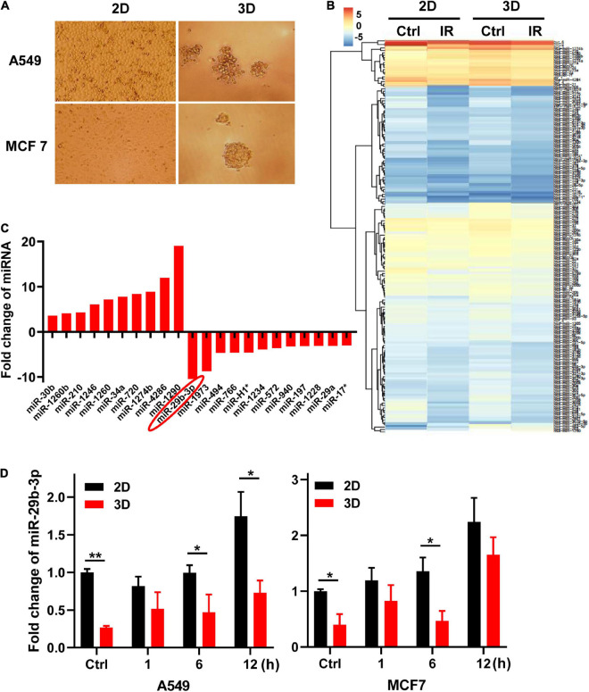
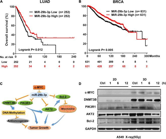
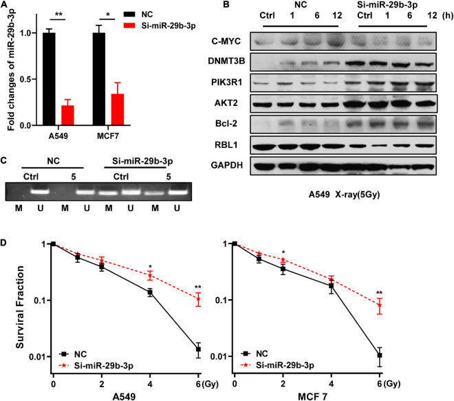
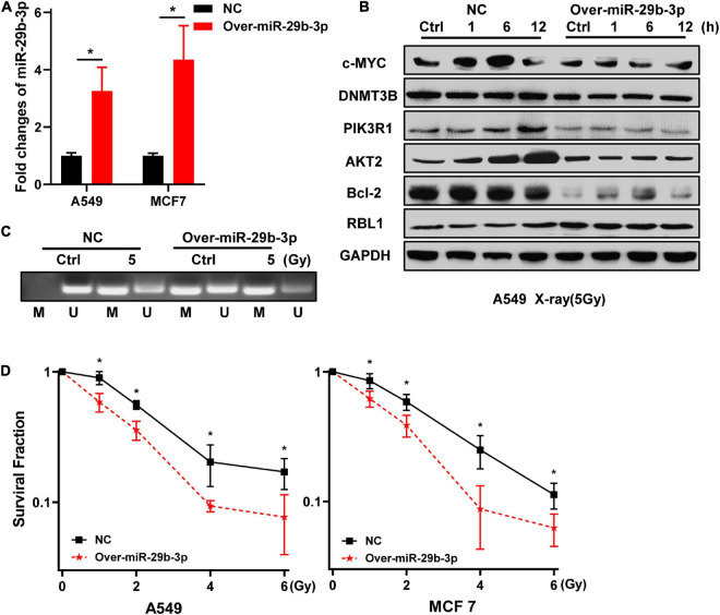
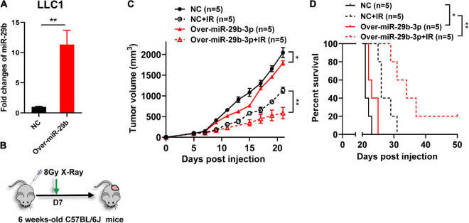
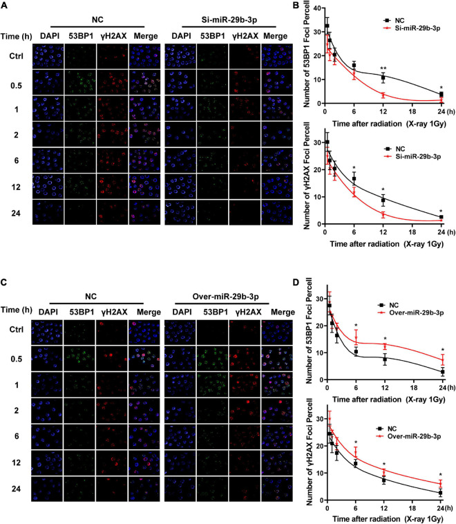
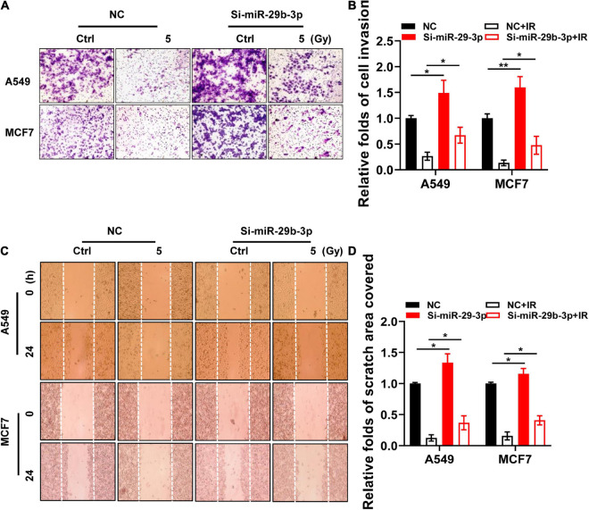
Radioresistance conferred by cancer stem cells (CSCs) is the principal cause of the failure of cancer radiotherapy. Eradication of CSCs is a prime therapeutic target and a requirement for effective radiotherapy. Three dimensional (3D) cell-cultured model could mimic the morphology of cells in vivo and induce CSC properties. Emerging evidence suggests that microRNAs (miRNAs) play crucial roles in the regulation of radiosensitivity in cancers. In this study, we aim to investigate the effects of miRNAs on the radiosensitivity of 3D cultured stem-like cells. Using miRNA microarray analysis in 2D and 3D cell culture models, we found that the expression of miR-29b-3p was downregulated in 3D cultured A549 and MCF7 cells compared with monolayer (2D) cells. Clinic data analysis from The Cancer Genome Atlas database exhibited that miR-29b-3p high expression showed significant advantages in lung adenocarcinoma and breast invasive carcinoma patients' prognosis. The subsequent experiments proved that miR-29b-3p overexpression decreased the radioresistance of cells in 3D culture and tumors in vivo through interfering kinetics process of DNA damage repair and inhibiting oncogenes RBL1, PIK3R1, AKT2, and Bcl-2. In addition, miR-29b-3p knockdown enhanced cancer cells invasion and migration capability. MiR-29b-3p overexpression decreased the stemness of 3D cultured cells. In conclusion, our results demonstrate that miR-29b-3p could be a sensitizer of radiation killing in CSC-like cells via inhibiting oncogenes expression. MiR-29b-3p could be a novel therapeutic candidate target for radiotherapy.

Keywords: miR-29b-3p; oncogene axis; radiosensitivity; stemness; three dimensional cultured cells.


Conflict of interest statement

The authors declare that the research was conducted in the absence of any commercial or financial relationships that could be construed as a potential conflict of interest.

Figures

FIGURE 1
FIGURE 1
MiR-29b-3p expression in 2D and 3D cultured A549 and MCF7 cells. (A) The morphology of 2D and 3D cultured A549 and MCF7 cells captured under a phase-contrast microscope. (B) Heat map of miRNAs microarray analysis in 2D and 3D cultured A549 cells 0.5 h after 5 Gy X-ray radiation. (C) Fold changes of miRNAs expression that are more than threefold between 2D and 3D cultured cells. (D) Relative miR-29b-3p expression measured by qRT-PCR at indicated time points in 2D and 3D cultured A549 and MCF7 cells after 5 Gy X-ray irradiation. U6 was used as an internal control. Ctrl, unirradiated groups. Significance was determined by unpaired t-test. *P < 0.05; **P < 0.01.
FIGURE 2
FIGURE 2
MiR-29b-3p expression affects survival probability in LUAD and BRCA by regulating oncogenes. (A) Kaplan–Meier survival curves of LUAD patients with low or high miR-29b-3p expression. (B) Kaplan–Meier survival curves of BRCA patients with low or high miR-29b-3p expression. (C) A schematic representation showing the signaling pathway of miR-29b-3p suppressing tumor growth by targeting a series of oncogenes. (D) Western blot assay on the expression of c-MYC, DNMT3B, PIK3R1, AKT2 and Bcl-2 at the indicated time points after 5 Gy X-rays in 2D and 3D cultured A549 cells. Ctrl, unirradiated groups. The cut-off values of high and low are median in LUAD and BRCA. P-values calculated by use of logrank test.
FIGURE 3
FIGURE 3
Knockdown of miR-29b-3p enhances the radioresistance of 2D cultured A549 and MCF7 cells by regulating oncogenes. (A) Relative expression levels of miR-29b-3p measured by qRT-PCR in 2D miR-29b-3p knockdown A549 and MCF7 cells. (B) Western blot assay on the expression of c-MYC, DNMT3B, PIK3R1, AKT2, Bcl-2, and RBL1 at indicated time points after 5 Gy X-rays in 2D cultured miR-29b-3p knockdown A549 cells. (C) The changes of the promoter methylation status of RBL1 measured by MSP in 5 Gy X-ray irradiated and unirradiated 2D cultured miR-29b-3p knockdown A549 cells. (D) Colony formation assay on the 2D cultured miR-29b-3p knockdown A549 and MCF7 cells after exposure to 0, 1, 2, 4 and 6 Gy X-rays. Ctrl, unirradiated groups. NC: negative control. Significance was determined by unpaired t-test. *P < 0.05; **P < 0.01.
FIGURE 4
FIGURE 4
Overexpression of miR-29b-3p sensitizes the 3D cultured A549 and MCF7 cells to radiation. (A) Relative expression of miR-29b-3p measured by qRT-PCR in 3D culture miR-29b-3p overexpressed A549 and MCF7 cells. (B) Western blot assay on the expression of c-MYC, DNMT3B, PIK3R1, AKT2, Bcl-2 and RBL1 at indicated time points after 5 Gy X-rays in 3D cultured miR-29b-3p overexpressed A549 cells. (C) The changes of the promoter methylation status of RBL1 measured by MSP in 5 Gy X-ray irradiated and unirradiated 3D cultured miR-29b-3p overexpressed A549 cells. (D) Colony formation assay on the 3D cultured miR-29b-3p overexpressed A549 and MCF7 cells after exposure to 0, 1, 2, 4 and 6 Gy X-rays. Ctrl, unirradiated groups; NC, negative control. Significance was determined by unpaired t-test. *P < 0.05.
FIGURE 5
FIGURE 5
The miR-29b-3p expression level on the efficacy of radiotherapy in vivo. (A) Relative expression of miR-29b-3p measured by qRT-PCR in miR-29b-3p overexpressed LLC1 cells. (B) Protocol for radiation treatment. (C,D) Tumor growth and Kaplan–Meier survival curves of C57BL/6 mice inoculated with 2 × 10⁵ VC or miR-29b-3p overexpressed LLC1 cells. Radiotherapy was conducted lx at 8 Gy at 7 days post inoculation of the tumor cells. NC, negative control. Significance was determined by 2-way ANOVA in c, logrank test in d respectively. *P < 0.05; **P < 0.01.
FIGURE 6
FIGURE 6
Kinetics process of DNA damage repair in miR-29b-3p knockdown or overexpressed A549 cells. (A) Graph shows quantification of 53BP1 and yH2AX foci in miR-29b-3p knockdown and negative control A549 cells exposed to 1 Gy X-ray. (B) The numbers of 53BP1 and yH2AX foci in 50 cells of each group were counted for each time point. (C) Graph shows quantification of 53BP1 and yH2AX foci in miR-29b-3p overexpressed and negative control A549 cells exposed to 1 Gy X-ray. (D) The numbers of 53BP1 and yH2AX foci in 50 cells of each group were counted for each time point. Ctrl, unirradiated groups; NC, negative control. Significance was determined by unpaired t-test. *P < 0.05; **P < 0.01.
FIGURE 7
FIGURE 7
Invasive and migratory capacity of irradiated nniR-29b-3p knockdown A549 and MCF7 cells. (A,B) Matrigel invasion assays in miR-29b-3p knockdown or negative control A549 and MCF7 cells after 5 Gy X-ray irradiation at 0 and 48 h. (C,D) Scratch wound healing migration assays in miR-29b-3p knockdown or negative control A549 and MCF7 cells after 5 Gy X-ray irradiation at 0 h and 24 h. Ctrl, unirradiated groups; NC, negative control. Significance was determined by unpaired t-test. *P < 0.05; **P < 0.01.
FIGURE 8
FIGURE 8
Correlation between miR-29b-3p expression and cancer cell sternness. (A) Sorting CD133+ A549 cells by flow cytometry. (B) Relative miR-29b-3p expression measured by qRT-PCR in CD133+ and negative control A549 cells. (C) Western blot assay on the expression of CD133, c-MYC, DNMT3B, PIK3R1, AKT2, Bcl-2 and RBL1 at indicated time points after 5 Gy X-rays in CD133+ A549 cells and negative control cells. (D,E) Side population in miR-29b-3p knockdown 2D and overexpressed 3D culture A549 cells. Ctrl, unirradiated groups; NC, negative control; WT, wild type groups. Significance was determined by unpaired t-test. *P < 0.05; **P < 0.01.

Similar articles

Cited by

References

    1. Asaithamby A., Hu B., Delgado O., Ding L. H., Story M. D., Minna J. D., et al. (2011). Irreparable complex DNA double-strand breaks induce chromosome breakage in organotypic three-dimensional human lung epithelial cell culture. Nucleic Acids Res. 39 5474–5488. 10.1093/nar/gkr149 - DOI - PMC - PubMed
    1. Ayob A. Z., Ramasamy T. S. (2018). Cancer stem cells as key drivers of tumour progression. J. Biomed. Sci. 25:20. 10.1186/s12929-018-0426-4 - DOI - PMC - PubMed
    1. Bartel D. P. (2004). MicroRNAs: genomics, biogenesis, mechanism, and function. Cell 116 281–297. - PubMed
    1. Berghauser Pont L. M. E., Spoor J. K. H., Venkatesan S., Swagemakers S., Kloezeman J. J., Dirven C. M., et al. (2014). The Bcl-2 inhibitor Obatoclax overcomes resistance to histone deacetylase inhibitors SAHA and LBH589 as radiosensitizers in patient-derived glioblastoma stem-like cells. Genes Cancer 5 445–459. 10.18632/genesandcancer.42 - DOI - PMC - PubMed
    1. Blazek E. R., Foutch J. L., Maki G. (2007). Daoy medulloblastoma cells that express CD133 are radioresistant relative to CD133- cells, and the CD133+ sector is enlarged by hypoxia. Int. J. Radiat. Oncol. Biol. Phys. 67 1–5. 10.1016/j.ijrobp.2006.09.037 - DOI - PubMed