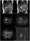
Attempt of Real-Time Near-Infrared Fluorescence Imaging Using Indocyanine Green (ICG) in Radical Resection of Gallbladder Cancer: A Case Report
- PMID: 34604291
- PMCID: PMC8481662
- DOI: 10.3389/fsurg.2021.655805
Attempt of Real-Time Near-Infrared Fluorescence Imaging Using Indocyanine Green (ICG) in Radical Resection of Gallbladder Cancer: A Case Report
Abstract
Surgery is the mainstay of treatment for resectable gallbladder cancer. Near-infrared fluorescence (NIRF) imaging using ICG is an innovation in laparoscopic surgery, which can provide real-time navigation during the whole operation. In this article, we present a 56-year older woman with gallbladder cancer, in which we evaluated the applicability of NIRF imaging using ICG for tumor and biliary tree visualization during the operative procedure of gallbladder cancer. The tumor and biliary tree were clearly visualized by utilizing a green fluorescence dye. The patient was successfully operated radical resection of gallbladder cancer under fluorescence laparoscope, without any complications. According to this case, the utilization of ICG based NIRF imaging is feasible and beneficial in identifying tumors and the biliary tree during radical resection. It can assist in the achievement of a negative margin and lymphatic clearance around the biliary tree. However, further studies are needed to corroborate the results of this case.
Keywords: case report; fluorescence laparoscope; gallbladder cancer; indocyanine green; near-infrared fluorescence.
Copyright © 2021 Yu, Xiang, Bai, Maswikiti, Gu, Li, Li, Zheng, Zhang and Chen.
Conflict of interest statement
The authors declare that the research was conducted in the absence of any commercial or financial relationships that could be construed as a potential conflict of interest.
Figures


Similar articles
-
Use of indocyanine green (ICG) augmented near-infrared fluorescence imaging in robotic radical resection of gallbladder adenocarcinomas.Surg Endosc. 2020 Jun;34(6):2490-2494. doi: 10.1007/s00464-019-07053-w. Epub 2019 Aug 6. Surg Endosc. 2020. PMID: 31388807
-
Near-infrared fluorescence cholangiography at a very low dose of indocyanine green: quantification of fluorescence intensity using a colour analysis software based on the RGB color model.Langenbecks Arch Surg. 2022 Dec;407(8):3513-3524. doi: 10.1007/s00423-022-02614-5. Epub 2022 Jul 26. Langenbecks Arch Surg. 2022. PMID: 35879621
-
Near-infrared cholecysto-cholangiography with indocyanine green may secure cholecystectomy in difficult clinical situations: proof of the concept in a porcine model.Surg Endosc. 2016 Sep;30(9):4115-23. doi: 10.1007/s00464-015-4608-9. Epub 2015 Oct 28. Surg Endosc. 2016. PMID: 26511116
-
Best practices in near-infrared fluorescence imaging with indocyanine green (NIRF/ICG)-guided robotic urologic surgery: a systematic review-based expert consensus.World J Urol. 2020 Apr;38(4):883-896. doi: 10.1007/s00345-019-02870-z. Epub 2019 Jul 8. World J Urol. 2020. PMID: 31286194
-
Systematic review of the role of indocyanine green near-infrared fluorescence in safe laparoscopic cholecystectomy (Review).Exp Ther Med. 2022 Feb;23(2):187. doi: 10.3892/etm.2021.11110. Epub 2021 Dec 30. Exp Ther Med. 2022. PMID: 35069868 Free PMC article. Review.
Cited by
-
Preoperative Assessment and Perioperative Management of Resectable Gallbladder Cancer in the Era of Precision Medicine and Novel Technologies: State of the Art and Future Perspectives.Diagnostics (Basel). 2022 Jul 5;12(7):1630. doi: 10.3390/diagnostics12071630. Diagnostics (Basel). 2022. PMID: 35885535 Free PMC article. Review.
References
-
- Eriksson AG, Beavis A, Soslow RA, Zhou Q, Abu-Rustum NR, Gardner GJ, et al. . A comparison of the detection of sentinel lymph nodes using indocyanine green and near-infrared fluorescence imaging versus blue dye during robotic surgery in uterine cancer. Int J Gynecol Cancer. (2017) 27:743–7. 10.1097/IGC.0000000000000959 - DOI - PMC - PubMed
-
- Frumovitz M, Plante M, Lee PS, Sandadi S, Lilja JF, Escobar PF, et al. . Near-infrared fluorescence for detection of sentinel lymph nodes in women with cervical and uterine cancers (FILM): a randomised, phase 3, multicentre, non-inferiority trial. Lancet Oncol. (2018) 19:1394–403. 10.1016/S1470-2045(18)30448-0 - DOI - PMC - PubMed
Publication types
LinkOut - more resources
Full Text Sources

