A Case Report of Calyceal Diverticulum: Differential Diagnosis for Organ-Preserving Operations
- PMID: 34604297
- PMCID: PMC8483267
- DOI: 10.3389/fsurg.2021.731796
A Case Report of Calyceal Diverticulum: Differential Diagnosis for Organ-Preserving Operations
Abstract
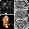
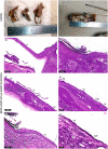
Calyceal diverticula and epidermal cysts are extremely rare kidney lesions with unknown etiology and pathogenesis. They have non-specific clinical and radiological picture. Despite the benign nature, sometimes these disorders mimic malignant tumors leading to unjustified nephrectomy. We present a clinical and morphological observation of a multicystic lesion in a 76-year-old patient's right kidney filled with keratinized masses and imitating a malignant solid tumor. The detailed gross, histological and immunohistochemical (desmin, cytokeratin 7, uroplakin and p63) analyses of the kidney tissue excluded the malignant nature of the lesion. The final differential diagnosis was between an epidermal cyst and calyceal diverticulum with pronounced squamous cell metaplasia of urothelium. The upper pole localization of the lesion, its connection with the pelvicalyceal system through the unobstructed isthmus, the presence of urothelial lining and smooth muscle cells in its wall let us diagnose a calyceal diverticulum type I. Knowledge of the key clinical and morphological features of epidermal cysts and diverticula of the pelvicalyceal system will help the practicing physicians suspect the benign nature of such lesions and perform organ-preserving operations.
Keywords: calyceal diverticulum; intrarenal epidermal cysts; kidney; nephrectomy; simple renal cyst.
Copyright © 2021 Kurkov, Pominalnaya, Nechay, Ratke, Mishugin, Drobyazko, Butenko, Fayzullin and Gomzikova.
Conflict of interest statement
The authors declare that the research was conducted in the absence of any commercial or financial relationships that could be construed as a potential conflict of interest.
Figures


Similar articles
-
Calyceal diverticulum of the kidney in pediatric patients - Is it as rare as you might think?J Pediatr Urol. 2020 Aug;16(4):487.e1-487.e6. doi: 10.1016/j.jpurol.2020.05.151. Epub 2020 May 29. J Pediatr Urol. 2020. PMID: 32580877
-
Type 2 calyceal diverticulum with an unusual appearance in the lower pole of the kidney.J Radiol Case Rep. 2022 Jun 30;16(6):12-17. doi: 10.3941/jrcr.v16i6.4334. eCollection 2022 Jun. J Radiol Case Rep. 2022. PMID: 35875367 Free PMC article.
-
Squamous Cell Carcinoma in a Calyceal Diverticulum Detected by Percutaneous Nephroscopic Biopsy.Case Rep Oncol Med. 2018 Jul 24;2018:3508537. doi: 10.1155/2018/3508537. eCollection 2018. Case Rep Oncol Med. 2018. PMID: 30140478 Free PMC article.
-
Challenges in the diagnosis of calyceal diverticulum: A report of two cases and review of the literature.J Xray Sci Technol. 2019;27(6):1155-1167. doi: 10.3233/XST-190549. J Xray Sci Technol. 2019. PMID: 31476195 Review.
-
Intrarenal epidermal cyst.Pathol Int. 2003 Aug;53(8):574-8. doi: 10.1046/j.1440-1827.2003.01512.x. Pathol Int. 2003. PMID: 12895239 Review.
Cited by
-
Prolonged postoperative urine leakage due to a calyceal diverticulum mimicking a renal cyst: A case report and literature review.Front Surg. 2022 Sep 7;9:967525. doi: 10.3389/fsurg.2022.967525. eCollection 2022. Front Surg. 2022. PMID: 36157402 Free PMC article.
-
The Pathophysiology of Inherited Renal Cystic Diseases.Genes (Basel). 2024 Jan 11;15(1):91. doi: 10.3390/genes15010091. Genes (Basel). 2024. PMID: 38254980 Free PMC article. Review.
References
Publication types
LinkOut - more resources
Full Text Sources
Research Materials

