

Identification of plasma proteins relating to brain neurodegeneration and vascular pathology in cognitively normal individuals
- PMID: 34604499
- PMCID: PMC8474123
- DOI: 10.1002/dad2.12240
Identification of plasma proteins relating to brain neurodegeneration and vascular pathology in cognitively normal individuals
Abstract
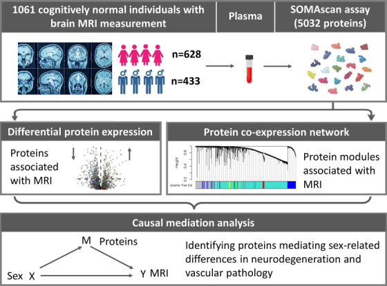
Introduction: This study aims to first discover plasma proteomic biomarkers relating to neurodegeneration (N) and vascular (V) damage in cognitively normal individuals and second to discover proteins mediating sex-related difference in N and V pathology.
Methods: Five thousand and thirty-two plasma proteins were measured in 1061 cognitively normal individuals (628 females and 433 males), nearly 90% of whom had magnetic resonance imaging measures of hippocampal volume (as N) and white matter hyperintensities (as V).
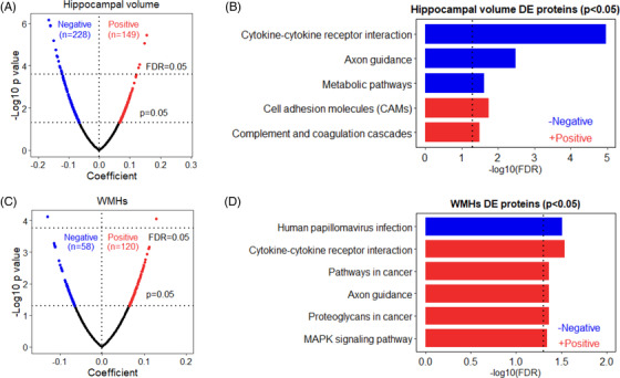
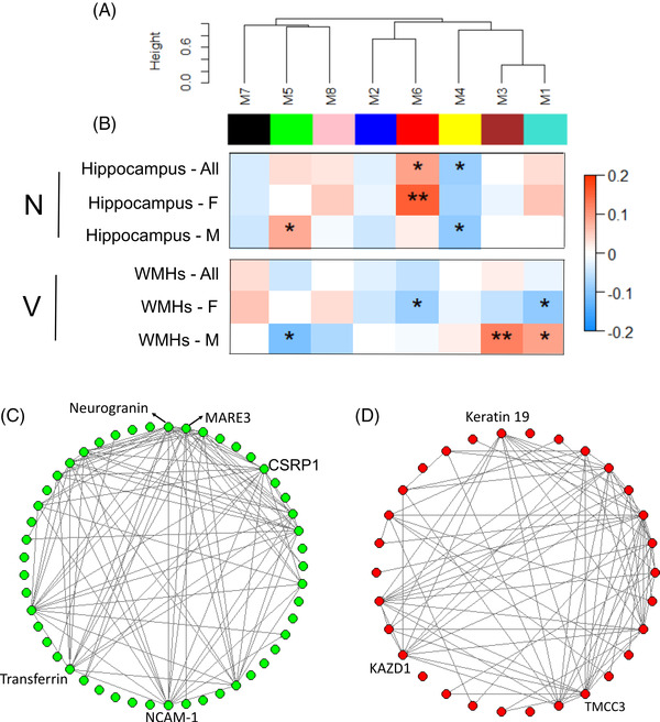
Results: Differential protein expression analysis and co-expression network analysis revealed different proteins and modules associated with N and V, respectively. Furthermore, causal mediation analysis revealed four proteins mediated sex-related difference in N and one protein mediated such difference in V damage.
Discussion: Once validated, the identified proteins could help to select cognitively normal individuals with N and V pathology for Alzheimer's disease clinical trials and provide targets for further mechanistic studies on brain sex differences, leading to sex-specific therapeutic strategies.
Keywords: mediation; neurodegeneration; plasma proteomics; sex‐related difference; vascular damage.
© 2021 The Authors. Alzheimer's & Dementia: Diagnosis, Assessment & Disease Monitoring published by Wiley Periodicals, LLC on behalf of Alzheimer's Association.
Conflict of interest statement
S.R.C received payment from the Society of Biological Psychiatry (plenary at SOBP2021). R.E.M. is an advisor to the Epigenetic Clock Development Foundation and has received a speaker fee from Illumina. A.C. is member of Edinburgh MVM Research Ethics Committee. J.M.W is involved in European Stroke Organisation Guideline on Covert Small Vessel Disease 2021 and European Stroke Organisation Chair of Conference Planning Group 2021 and 2022. D.S. helped to set up CAPE study. A.M. received speaker fees from Janssen and Illumina. S.L. is an employee of Janssen Medical UK and Co‐founder of Akrivia Health Ltd. He is also named as an inventor on biomarker intellectual property protected by Proteome Sciences and Kings College London unrelated to the current study and within the past 5 years has advised for Optum labs, Merck, SomaLogic, and been the recipient of funding from AstraZeneca and other companies via the IMI funding scheme. N.B. is a member of Mehta Family Centre for Engineering in Medicine, Kanpur, India as well as the editorial board of Stem Cells. A.N.H. is the main PI of a project funded by J&J, and another projects funded by GSK, all unrelated to this study. The remaining authors declare that the research was conducted in the absence of any commercial or financial relationships that could be construed as a potential conflict of interest.
Figures
Similar articles
-
Stage-specific links between plasma neurofilament light and imaging biomarkers of Alzheimer's disease.Brain. 2020 Dec 1;143(12):3793-3804. doi: 10.1093/brain/awaa342. Brain. 2020. PMID: 33210117 Free PMC article.
-
Cerebrovascular disease promotes tau pathology in Alzheimer's disease.Brain Commun. 2020 Aug 19;2(2):fcaa132. doi: 10.1093/braincomms/fcaa132. eCollection 2020. Brain Commun. 2020. PMID: 33215083 Free PMC article.
-
Plasma Markers of Alzheimer's Disease Pathology, Neuronal Injury, and Astrocytic Activation and MRI Load of Vascular Pathology and Neurodegeneration: The SMART-MR Study.J Am Heart Assoc. 2024 Feb 20;13(4):e032134. doi: 10.1161/JAHA.123.032134. Epub 2024 Feb 14. J Am Heart Assoc. 2024. PMID: 38353228 Free PMC article.
-
Sex differences in functional and molecular neuroimaging biomarkers of Alzheimer's disease in cognitively normal older adults with subjective memory complaints.Alzheimers Dement. 2018 Sep;14(9):1204-1215. doi: 10.1016/j.jalz.2018.05.014. Epub 2018 Jul 7. Alzheimers Dement. 2018. PMID: 30201102 Review.
-
White matter hyperintensities are higher among early-onset Alzheimer's disease participants than their cognitively normal and early-onset nonAD peers: Longitudinal Early-onset Alzheimer's Disease Study (LEADS).Alzheimers Dement. 2023 Nov;19 Suppl 9(Suppl 9):S89-S97. doi: 10.1002/alz.13402. Epub 2023 Jul 25. Alzheimers Dement. 2023. PMID: 37491599 Free PMC article. Review.
Cited by
-
Blood-Based Proteomic Profiling Identifies Potential Biomarker Candidates and Pathogenic Pathways in Dementia.Int J Mol Sci. 2023 May 1;24(9):8117. doi: 10.3390/ijms24098117. Int J Mol Sci. 2023. PMID: 37175824 Free PMC article.
-
Identification and replication of sex-dimorphic protein quantitative trait loci across multiple ancestries and their associations with diseases.Sci Rep. 2025 Aug 28;15(1):31721. doi: 10.1038/s41598-025-10031-z. Sci Rep. 2025. PMID: 40877285 Free PMC article.
-
Identification of blood plasma protein ratios for distinguishing Alzheimer's disease from healthy controls using machine learning.Heliyon. 2025 Jan 28;11(3):e42349. doi: 10.1016/j.heliyon.2025.e42349. eCollection 2025 Feb 15. Heliyon. 2025. PMID: 39981365 Free PMC article.
-
Associations of plasma proteomics and age-related outcomes with brain age in a diverse cohort.Geroscience. 2024 Aug;46(4):3861-3873. doi: 10.1007/s11357-024-01112-4. Epub 2024 Mar 4. Geroscience. 2024. PMID: 38438772 Free PMC article.
-
Multi Layered Omics Approaches Reveal Glia Specific Alterations in Alzheimer's Disease: A Systematic Review and Future Prospects.Glia. 2025 Mar;73(3):539-573. doi: 10.1002/glia.24652. Epub 2024 Dec 9. Glia. 2025. PMID: 39652363 Free PMC article.
References
-
- Schröder J, Pantel J. Neuroimaging of hippocampal atrophy in early recognition of Alzheimer's disease–a critical appraisal after two decades of research. Psychiatry Res Neuroimaging. 2016;247:71‐78. - PubMed
-
- Seab JP, Jagust WJ, Wong ST, Roos MS, Reed BR, Budinger TF. Quantitative NMR measurements of hippocampal atrophy in Alzheimer's disease. Magn Reson Med. 1988;8:200‐208. - PubMed
Grants and funding
LinkOut - more resources
Full Text Sources