Deep learning analysis of resting electrocardiograms for the detection of myocardial dysfunction, hypertrophy, and ischaemia: a systematic review
- PMID: 34604757
- PMCID: PMC8482047
- DOI: 10.1093/ehjdh/ztab048
Deep learning analysis of resting electrocardiograms for the detection of myocardial dysfunction, hypertrophy, and ischaemia: a systematic review
Erratum in
-
Erratum.Eur Heart J Digit Health. 2021 Nov 21;3(1):115-116. doi: 10.1093/ehjdh/ztab098. eCollection 2022 Mar. Eur Heart J Digit Health. 2021. PMID: 36713986 Free PMC article.
Abstract
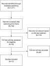
The aim of this review was to assess the evidence for deep learning (DL) analysis of resting electrocardiograms (ECGs) to predict structural cardiac pathologies such as left ventricular (LV) systolic dysfunction, myocardial hypertrophy, and ischaemic heart disease. A systematic literature search was conducted to identify published original articles on end-to-end DL analysis of resting ECG signals for the detection of structural cardiac pathologies. Studies were excluded if the ECG was acquired by ambulatory, stress, intracardiac, or implantable devices, and if the pathology of interest was arrhythmic in nature. After duplicate reviewers screened search results, 12 articles met the inclusion criteria and were included. Three articles used DL to detect LV systolic dysfunction, achieving an area under the curve (AUC) of 0.89-0.93 and an accuracy of 98%. One study used DL to detect LV hypertrophy, achieving an AUC of 0.87 and an accuracy of 87%. Six articles used DL to detect acute myocardial infarction, achieving an AUC of 0.88-1.00 and an accuracy of 83-99.9%. Two articles used DL to detect stable ischaemic heart disease, achieving an accuracy of 95-99.9%. Deep learning models, particularly those that used convolutional neural networks, outperformed rules-based models and other machine learning models. Deep learning is a promising technique to analyse resting ECG signals for the detection of structural cardiac pathologies, which has clinical applicability for more effective screening of asymptomatic populations and expedited diagnostic work-up of symptomatic patients at risk for cardiovascular disease.
Keywords: Artificial intelligence; Coronary artery disease; Deep learning; Electrocardiogram; Heart failure; Left ventricular hypertrophy; Myocardial infarction.
© The Author(s) 2021. Published by Oxford University Press on behalf of the European Society of Cardiology.
Figures


References
-
- Schlant RC, Adolph RJ, DiMarco JP, Dreifus LS, Dunn MI, Fisch C, et al. Guidelines for electrocardiography. A report of the American College of Cardiology/American Heart Association Task Force on Assessment of Diagnostic and Therapeutic Cardiovascular Procedures (Committee on Electrocardiography). Circulation 1992;85:1221–1228. - PubMed
-
- Sansone M, Fusco R, Pepino A, Sansone C.. Electrocardiogram pattern recognition and analysis based on artificial neural networks and support vector machines: a review. J Healthc Eng 2013;4:465–504. - PubMed
-
- Guglin ME, Thatai D.. Common errors in computer electrocardiogram interpretation. Int J Cardiol. 2006;106:232–237. - PubMed
-
- Poon K, Okin PM, Kligfield P.. Diagnostic performance of a computer-based ECG rhythm algorithm. J Electrocardiol 2005;38:235–238. - PubMed
Publication types
LinkOut - more resources
Full Text Sources
Miscellaneous
