. 2021 Jul 26;1(9):1368-1379.
doi: 10.1021/jacsau.1c00173. eCollection 2021 Sep 27.

Toward Point-of-Care Detection of Mycobacterium tuberculosis: A Brighter Solvatochromic Probe Detects Mycobacteria within Minutes

Affiliations

Toward Point-of-Care Detection of Mycobacterium tuberculosis: A Brighter Solvatochromic Probe Detects Mycobacteria within Minutes

Mireille Kamariza et al. JACS Au. .

Abstract

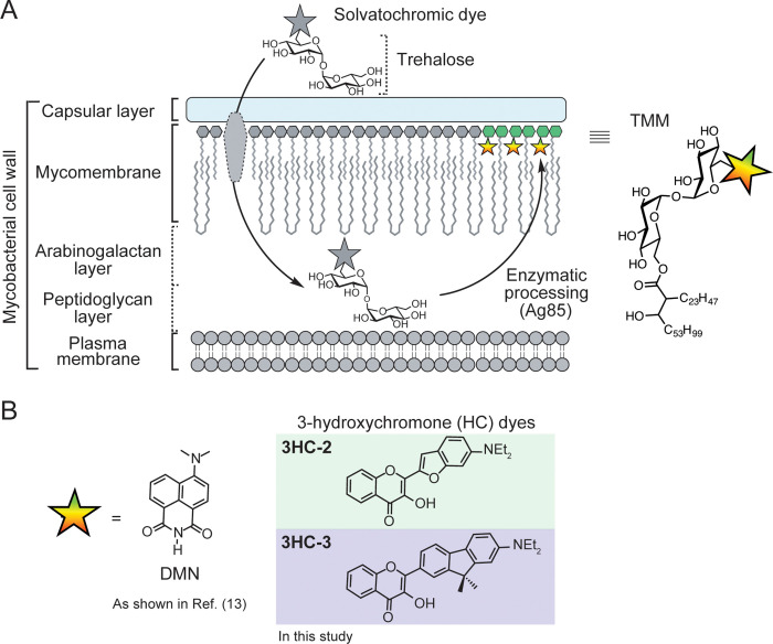
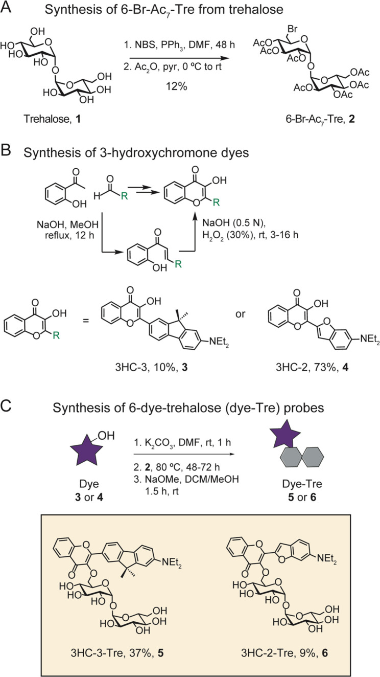
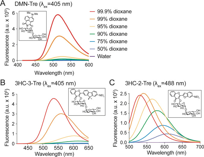
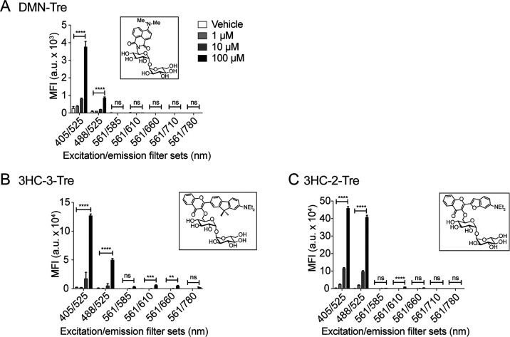
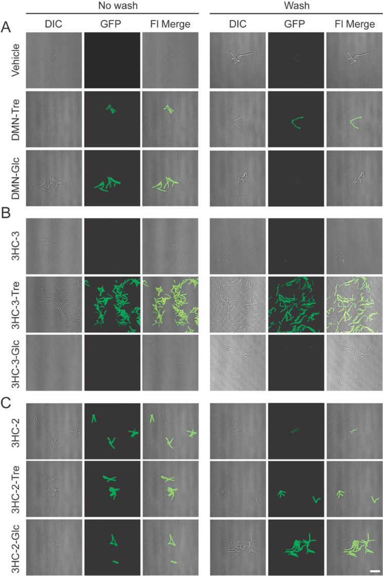
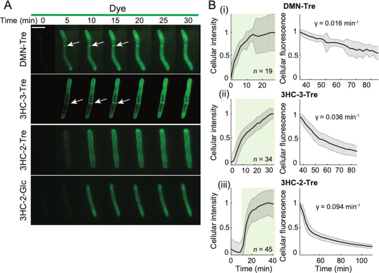
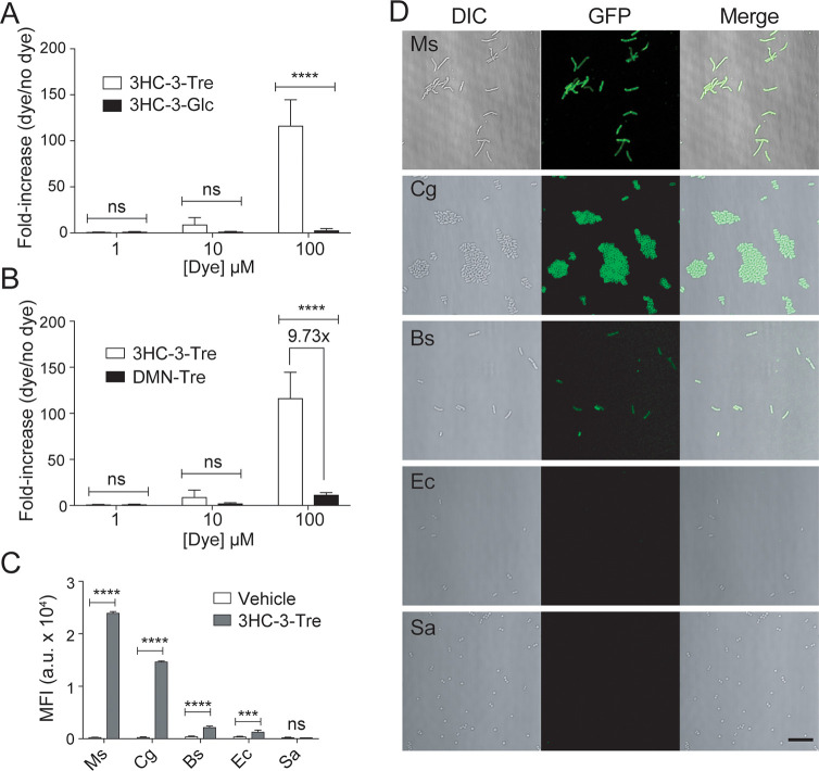
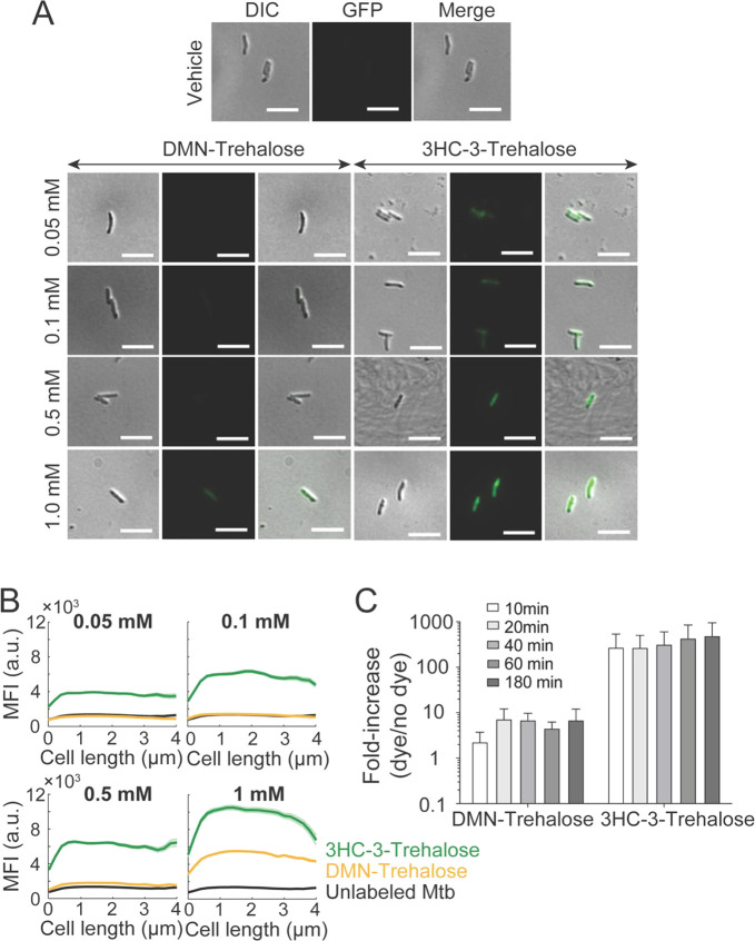
There is an urgent need for point-of-care tuberculosis (TB) diagnostic methods that are fast, inexpensive, and operationally simple. Here, we report on a bright solvatochromic dye trehalose conjugate that specifically detects Mycobacterium tuberculosis (Mtb) in minutes. 3-Hydroxychromone (3HC) dyes, known for having high fluorescence quantum yields, exhibit shifts in fluorescence intensity in response to changes in environmental polarity. We synthesized two analogs of 3HC-trehalose conjugates (3HC-2-Tre and 3HC-3-Tre) and determined that 3HC-3-Tre has exceptionally favorable properties for Mtb detection. 3HC-3-Tre-labeled mycobacterial cells displayed a 10-fold increase in fluorescence intensity compared to our previous reports on the dye 4,4-N,N-dimethylaminonapthalimide (DMN-Tre). Excitingly, we detected fluorescent Mtb cells within 10 min of probe treatment. Thus, 3HC-3-Tre permits rapid visualization of mycobacteria that ultimately could empower improved Mtb detection at the point-of-care in low-resource settings.


Conflict of interest statement

The authors declare the following competing financial interest(s): M.K. and C.R.B. are cofounders of OliLux Biosciences which has licensed patents related to the work in this paper. B.K. is a scientific advisor to OliLux Biosciences. B.K is cofounder of SmartSpot Quality CC and scientific advisor to SmartSpot Quality CC and Pulmosat. C.R.B. is a cofounder and scientific advisory board member of Lycia Therapeutics, Palleon Pharmaceuticals, Enable Bioscience, Redwood Biosciences (a subsidiary of Catalent), and InterVenn Biosciences, and a member of the board of directors of Eli Lilly & Company.

Figures

Figure 1
Figure 1
Solvatochromic trehalose probes label the mycobacterial mycomembrane. (A) Solvatochromic trehalose probes are converted by mycobacteria to the corresponding trehalose monomycolate (TMM, structure on right) analogs and inserted into the mycomembrane. There, they undergo fluorescence turn-on, enabling detection of labeled cells by fluorescence microscopy. (B) Chemical structures of solvatochromic dyes described in this study.
Figure 2
Figure 2
Synthesis scheme for 3-hydroxychromone (3HC) trehalose (Tre) dye conjugates.
Figure 3
Figure 3
Emission spectra of 3-hydroxychromone trehalose dyes. Fluorescence spectra of (A) DMN-Tre (ex. 405 nm), (B) 3HC-3-Tre (ex. 405 nm), and (C) 3-HC-2-Tre (ex. 488 nm) in solvent systems with the indicated ratios of dioxane in water.
Figure 4
Figure 4
Flow cytometry analysis of Msmeg cells labeled with solvatochromic trehalose dyes using various excitation and emission filter sets. Flow cytometry analysis of Msmeg labeled with (A) DMN-Tre, (B) 3HC-3-Tre, or (C) 3HC-2-Tre. All dyes showed increased labeling at the highest concentration (100 μM). Cells at OD600 = 0.5 were incubated with the indicated dye-trehalose probe concentrations for 1 h at 37 °C. MFI, mean fluorescence intensity. Data are means ± SEM from at least two independent experiments. Data were analyzed by two-way ANOVA tests for unequal variances with Dunn’s multiple comparisons test using selected adjusted P values. (*, p < 0.05; **, p < 0.01; ***, p < 0.001; ****, p < 0.0001; ns, not significant).
Figure 5
Figure 5
Unlike 3HC-2-Tre, 3HC-3-Tre labeling is dependent on the trehalose moeity. Epifluorescence microscopy of Msmeg cells treated with (A) 100 μM DMN-Tre or DMN-Glc or no dye control (Vehicle); (B) 100 μM of 3HC-3, 3HC-3-Tre, or 3HC-3-Glc; (C) 100 μM of 3HC-2, 3HC-2-Tre, or 3HC-2-Glc. 3HC-3-Tre showed the most efficient labeling of Msmeg. Cells were incubated with the indicated dyes for 1 h at 37 °C. Cells were smeared directly (No wash) or washed 3 times with PBS then smeared onto a microscope slide (Wash). Scale bar: 10 μm.
Figure 6
Figure 6
Unlike 3HC-2 dye conjugates, 3HC-3-Tre labeling is initially localized at the septum and poles. (A) Time-lapse microscopy of Msmeg cells treated with 100 μM DMN-Tre, 3HC-3-Tre, 3HC-2-Tre, or 3HC-2-Glc for 30 min revealed concentration of 3HC-3-Tre at cell septa and poles. White arrows denote septal labeling. Scale bar: 5 μm. (B) Quantification of Msmeg fluorescence in the presence of 100 μM (i) DMN-Tre, (ii) 3HC-3-Tre, or (iii) 3HC-2-Tre during labeling for 30 min (left, volume-normalized intensity) and subsequent washing with growth medium for 1 h (right, total fluorescence). The number of cells included in each analysis (n) is provided in each panel. Shaded error bars represent ±1 standard deviation.
Figure 7
Figure 7
Specific labeling of live mycobacteria and corynebacteria with 3HC-3-Tre. (A,B) Flow cytometry analysis of Msmeg cells incubated for 1 h at 37 °C with (A) 100 μM 3HC-3-Tre or 3HC-3-Glc, (B) 100 μM 3HC-3-Tre or DMN-Tre. (C,D) Flow cytometry (C) and no-wash microscopy (D) analyses of Msmeg (Ms), C. glutamicum (Cg), B. subtilis (Bs), E. coli (Ec), and S. aureus (Sa) cells incubated for 1 h at 37 °C with 100 μM 3HC-3-Tre. 3HC-3-Tre labeling was specific to Msmeg and Cg. Data are means ± SEM from at least two independent experiments. Data were analyzed by two-way ANOVA tests for unequal variances with Dunn’s multiple comparisons test using selected adjusted p-values (***, p < 0.001; ****, p < 0.0001; ns, not significant). Scale bar: 10 μm.
Figure 8
Figure 8
Mtb cells labeled with 3HC-3-Tre exhibit increased fluorescence intensities and can be detected within 10 min by flow cytometry. (A,B) Log-phase Mtb RvS cells (OD600nm ∼ 0.5) were stained with various concentrations of DMN-Tre or 3HC-3-Tre (0.05, 0.1, 0.5, and 1 mM) for 3 h at 37 °C. The vehicle sample served as the unlabeled control. Mtb cells were then fixed in 2.5% glutaraldehyde for 1.5 h at 37 °C, washed twice with 1X PBS, and re-suspended in 100 μL 1X PBS. Ten microliters of each sample were spotted onto a 2% agarose pad and imaged with an epifluorescence microscope. Images are representative of each staining condition; scale bar represents 5 μm. Cells were then subjected to (A) microscopy analysis and (B) quantitative cell staining analysis (n = 85 cells for each condition). (C) Flow cytometry analysis of H37Rv Mtb cells labeled with 100 μM DMN-Tre or 3HC-3-Tre for 10, 20, 40, 60, and 180 min. Log-phase cultures (OD600 nm ∼ 0.5) were stained with trehalose probes for the indicated times and then cell fluorescence was measured by flow cytometry. The fluorescence fold-increase (dye/nodye) was calculated as follows: fold-increase (dye/nodye) = mean fluorescence intensity of stained cells over the unlabeled sample. Data are means ± SEM from three independent biological experiments. Data were analyzed by two-way ANOVA test (ns: not significant).

References

    1. World Health Organization . Global Tuberculosis Report 2019; World Health Organization; S.l., 2019.
    1. World Health Organization . High-Priority Target Product Profiles for New Tuberculosis Diagnostics: Report of a Consensus Meeting; Geneva, Switzerland, 2014.
    1. Singhal R.; Myneedu V. P. Microscopy as a Diagnostic Tool in Pulmonary Tuberculosis. International Journal of Mycobacteriology 2015, 4 (1), 1–6. 10.1016/j.ijmyco.2014.12.006. - DOI - PubMed
    1. Ziehl F. Zur Farbung Des Tuberkelbacillus. Dtsch. Med. Wochenschr. 1882, 8, 451.10.1055/s-0029-1196721. - DOI
    1. Ziehl F. Ueber Die Farbung Des Tuberkelbacillus. Dtsch. Med. Wochenschr. 1883, 9, 247–249. 10.1055/s-0029-1197158. - DOI