. 2021 Oct;20(10):e13491.
doi: 10.1111/acel.13491. Epub 2021 Oct 3.

Advanced Maternal Age-associated SIRT1 Deficiency Compromises Trophoblast Epithelial-Mesenchymal Transition through an Increase in Vimentin Acetylation

Affiliations

Advanced Maternal Age-associated SIRT1 Deficiency Compromises Trophoblast Epithelial-Mesenchymal Transition through an Increase in Vimentin Acetylation

Liling Xiong et al. Aging Cell. 2021 Oct.

Abstract

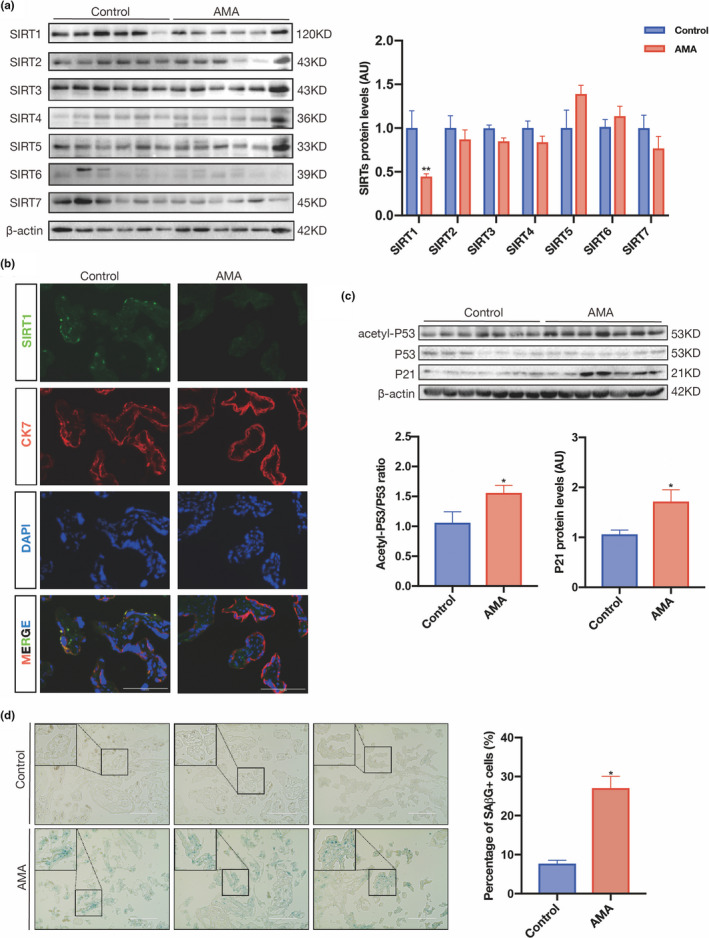
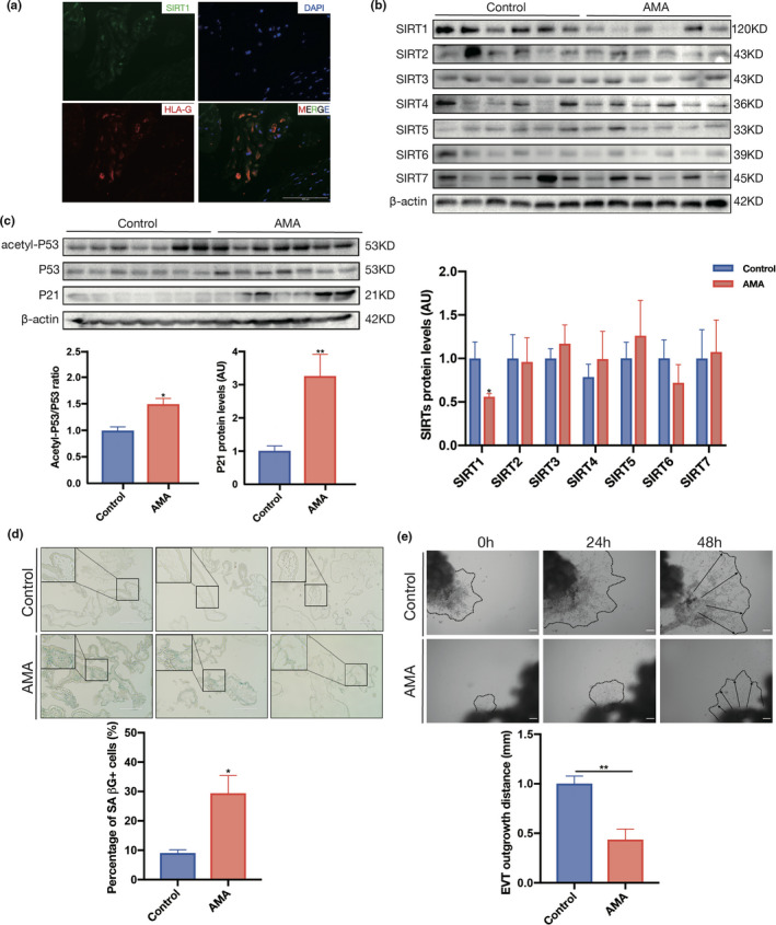
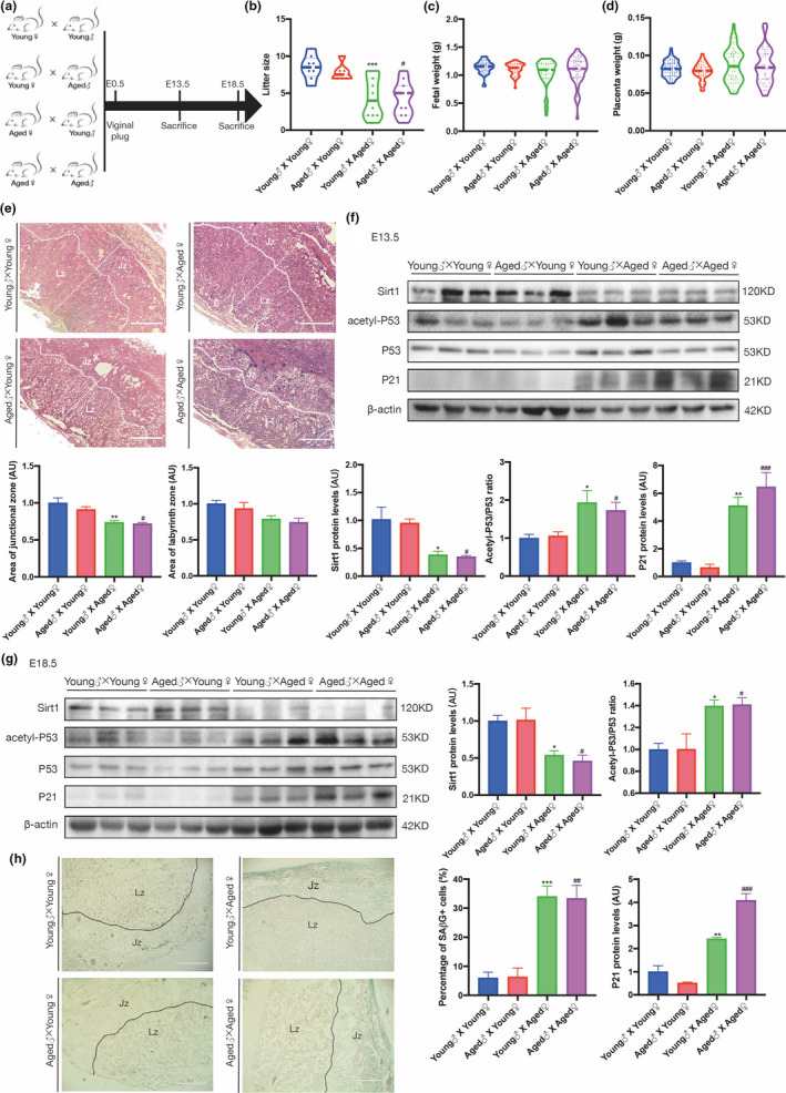
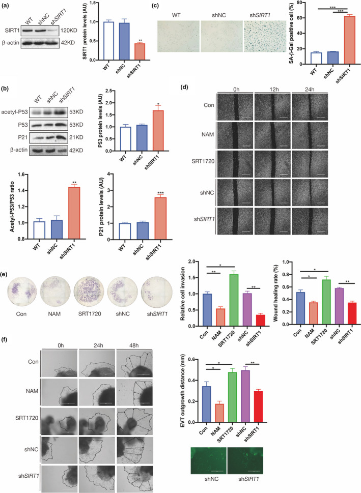
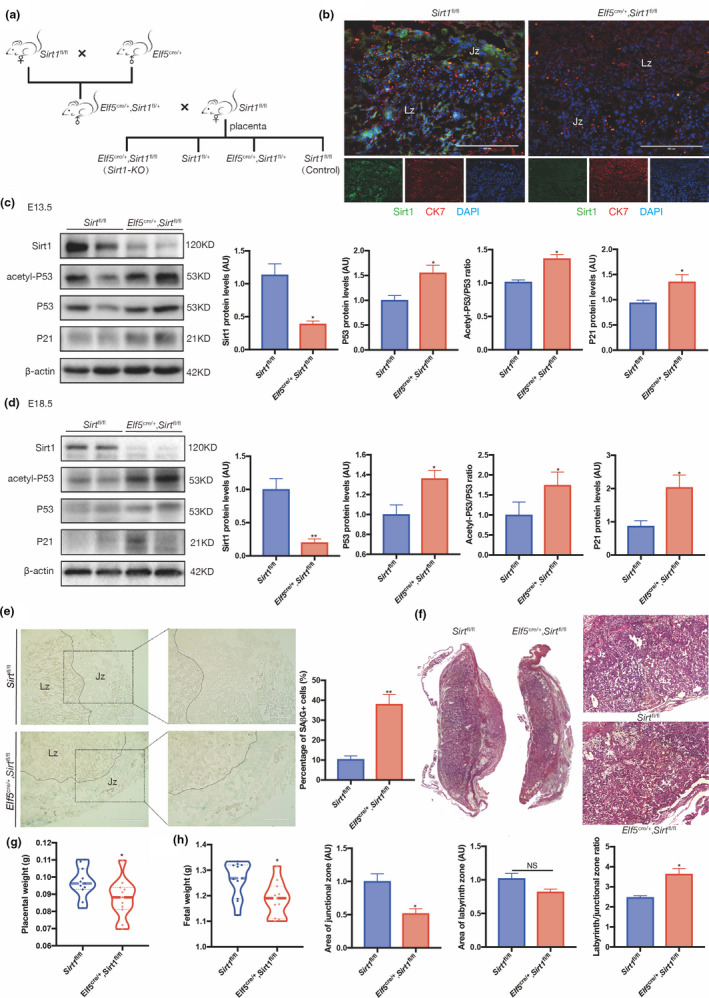
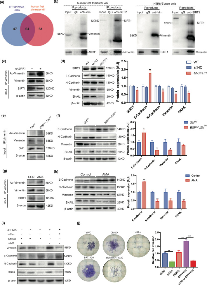
Advanced maternal age (AMA) pregnancies are rapidly increasing and are associated with aberrant trophoblast cell function, poor placentation, and unfavorable pregnancy outcomes, presumably due to premature placental senescence. SIRT1 is an NAD+ -dependent deacetylase with well-known antiaging effects, but its connection with placental senescence is unreported. In this study, human term placentas and first-trimester villi were collected from AMA and normal pregnancies, and a mouse AMA model was established by cross breeding young and aged male and female C57 mice. SIRT1 expression and activity in HTR8/SVneo cells were genetically or pharmacologically manipulated. Trophoblast-specific Sirt1-knockout (KO) mouse placentas were generated by mating Elf5-Cre and Sirt1fl/fl mice. Trophoblast cell mobility was assessed with transwell invasion and wound-healing assays. SIRT1-binding proteins in HTR8/SVneo cells and human placental tissue were identified by mass spectrometry. We identified SIRT1 as the only differentially expressed sirtuin between AMA and normal placentas. It is downregulated in AMA placentas early in the placental life cycle and is barely impacted by paternal age. SIRT1 loss upregulates P53 acetylation and P21 expression and impairs trophoblast invasion and migration. Sirt1-KO mouse placentas exhibit senescence markers and morphological disruption, along with decreased fetal weight. In trophoblasts, SIRT1 interacts with vimentin, regulating its acetylation. In conclusion, SIRT1 promotes trophoblast epithelial-mesenchymal transition (EMT) to enhance invasiveness by modulating vimentin acetylation. AMA placentas are associated with premature senescence during placentation due to SIRT1 loss. Therefore, SIRT1 may be an antiaging therapeutic target for improving placental development and perinatal outcomes in AMA pregnancies.

Keywords: SIRT1; advanced maternal age; epithelial−mesenchymal transition; placenta; senescence; trophoblast.


Conflict of interest statement

The authors have no conflicts of interest to declare.

Figures

FIGURE 1
FIGURE 1
SIRT1 is downregulated in AMA term placenta and is associated with placental senescence. (a) Western blot analysis of the expression of SIRT1–7 in human term placental tissues (n = 6 in the control group and AMA group). (b) IF staining of SIRT1 (green) and CK7 (red) in frozen sections of human term placentas; nuclei were counterstained with DAPI (blue). Scale bar: 100 μm. (c) Western blot analysis of the levels of acetyl‐P53, total P53, and P21 in human term placentas (n = 6 in the control group and AMA group). (d) SAβG staining of human term placental sections. Scale bar: 200 μm. AU, arbitrary unit. Data were analyzed using Student's t‐test, *p < 0.05. All data are presented as the mean ± SEM. All experiments were performed in triplicate
FIGURE 2
FIGURE 2
SIRT1 decreases in early AMA pregnancy and is associated with premature placental senescence. (a) IF detection of SIRT1 expression in first‐trimester human decidua. IF staining with an antibody against SIRT1 is shown in green, HLA‐G staining is shown in red, and nuclei are shown in blue (DAPI staining). (b) The expression of SIRT1–7 in human first‐trimester villi was determined using Western blotting (n = 6 in the control group and AMA group). (c) Western blot analysis of the levels of acetyl‐P53, P53, and P21 in human first‐trimester villi (n = 6 in the control group and AMA group). (d) SAβG staining of human first‐trimester villi sections. Scale bar: 200 μm. (e) Migration (dotted line circled area) of EVTs induced from human first‐trimester villous explants. n = 3; scale bar: 200 μm; AU, arbitrary unit. Data were analyzed using Student's t‐test, *p < 0.05, **p < 0.01. All data are presented as the mean ± SEM. All experiments were performed in triplicate
FIGURE 3
FIGURE 3
Advanced maternal age is associated with adverse birth outcomes and downregulation of SIRT1 in the mouse placenta. (a) The mating plan for the animals. (b) Average litter size. (c) Birth weight of viable fetuses. (d) Placental weight of viable fetuses among different mating groups. (e) H&E staining of sections from E13.5 placentas from different mating groups. Jz, junctional zone; Lz, labyrinth zone. Scale bar: 400 μm. Western blot analysis of the levels of SIRT1, acetyl‐P53, P53, and P21 in (f) E13.5 and (g) E18.5 mouse placentas from different mating groups (n = 3 biologically independent animals from each group). (h) SAβG staining of E13.5 mouse placental sections from different mating groups. Jz, junctional zone; Lz, labyrinth zone. Scale bar: 400 μm. AU, arbitrary unit. Data were analyzed using one‐way ANOVA, followed by Tukey's or Sidak's multiple comparison tests, n = 6; *p < 0.05; **p < 0.01; ***p < 0.001 vs. Young♂ x Young♀ group; #p < 0.05; ##p < 0.01; ###p < 0.001 vs. Aged♂ x Young♀ group. All data are presented as the mean ± SEM. All experiments were performed in triplicate
FIGURE 4
FIGURE 4
SIRT1 deficiency induces senescence and impairs the migration and invasion of trophoblast cell lines. (a) Western blot analysis of SIRT1 expression in HTR8/SVneo cells. (b) Western blot analysis of acetyl‐P53, P53, and P21 in HTR8/SVneo cells. (c) SAβG staining of HTR8/SVneo cells. Scale bar: 200 μm. (d) Wound‐healing assay; scale bar: 100 μm. (e) Transwell assay of HTR8/SVneo cells treated with SRT1720 or nicotinamide (NAM) and transfected with shSIRT1. (f) Migration (dotted line circled area) of EVTs induced from human first‐trimester villous explants treated with shSIRT1, SRT1720 and NAM. Scale bar: 200 μm. Data were analyzed using one‐way ANOVA; *p < 0.05, **p < 0.01, ***p < 0.001. All data are presented as the mean ± SEM. All experiments were performed in triplicate
FIGURE 5
FIGURE 5
Senescence and maldevelopment in the Sirt1‐deficient mouse placenta. (a) The mating plan for generating Sirt1‐deficient mouse placentas. (b) IF staining of Sirt1 (green), CK7 (red) and DAPI (blue) in E13.5 Elf5 cre/+, Sirt1 fl/fl and Sirt1 fl/fl mouse placental sections. Western blot analysis of SIRT1, acetyl‐P53, P53, and P21 levels in (c) E13.5 and (d) E18.5 mouse placentas (n = 3 biologically independent animals from each group). (e) SAβG staining of E13.5 mouse placenta from Elf5 cre/+, Sirt1 fl/fl and Sirt1 fl/fl mice; scale bar: 400 μm. (f) H&E staining of sections from Elf5 cre/+, Sirt1 fl/fl and Sirt1 fl/fl placentas collected on E18.5. Weights of (g) the Elf5 cre/+, Sirt1 fl/fl and Sirt1 fl/fl placentas and (h) the corresponding fetus on E18.5 are shown; scale bars, 400 μm. AU, arbitrary unit. Data were analyzed using Student's t‐test, n = 6, *p < 0.05, **p < 0.01. All data are presented as the mean ± SEM. All experiments were performed in triplicate
FIGURE 6
FIGURE 6
SIRT1 promotes trophoblast EMT. (a) Venn diagram of SIRT1‐binding proteins in HTR8/SVneo cells and human first‐trimester villi profiled by IP‐MS. (b) Reciprocal Co‐IP of SIRT1 and vimentin in human first‐trimester villi (left panel) and HTR8/SVneo cells (right panel). (c) Acetylation of vimentin in WT and shSIRT1 HTR8/SVneo cells. (d) Western blot analysis of E‐cadherin, N‐cadherin, vimentin, and SNAIL in WT, shNC, and shSIRT1 HTR8/SVneo cells. (e) Acetylation of vimentin in Elf5 cre/+, Sirt1 fl/fl and Sirt1 fl/fl mouse placentas collected on E13.5. (f) Western blot analysis of E‐cadherin, N‐cadherin, vimentin, and SNAIL levels in Elf5 cre/+, Sirt1 fl/fl and Sirt1 fl/fl mouse placentas collected on E13.5; n = 3. (g) Acetylation of vimentin in first‐trimester villi of AMA and control pregnancies. (h) Western blot analysis of E‐cadherin, N‐cadherin, vimentin, and SNAIL levels in first‐trimester villi from women with AMA and control women; n = 3. AU, arbitrary unit. Data were analyzed using Student's t‐test; *p < 0.05, **p < 0.01. All data are presented as the mean ± SEM. All experiments were performed in triplicate. (i) Transwell assays of Htr8/SVneo cells were performed after 48 h of treatment with siVim, SRT1720 or both. (j) Western blot analysis of E‐cadherin, N‐cadherin, vimentin, and SNAIL levels in Htr8/SVneo cells treated with siVim, SRT1720, or both for 48 h. Data were analyzed using one‐way ANOVA; **p < 0.01, ***p < 0.001. All data are expressed as the mean ± SEM

References

    1. Abdelmohsen, K. , Pullmann, R. Jr , Lal, A. , Kim, H. H. , Galban, S. , Yang, X. , Blethrow, J. , Walker, M. , Shubert, J. , Gillespie, D. A. , Furneaux, H. , & Gorospe, M. (2007). Phosphorylation of HuR by Chk2 regulates SIRT1 expression. Molecular Cell, 25(4), 543–557. - PMC - PubMed
    1. Abel, E. L. , Kruger, M. , & Burd, L. (2002). Effects of maternal and paternal age on Caucasian and native American preterm births and birth weights. American Journal of Perinatology, 19(01), 049–054. 10.1055/s-2002-20173 - DOI - PubMed
    1. Alves‐Fernandes, D. K. , & Jasiulionis, M. G. (2019). The role of SIRT1 on DNA damage response and epigenetic alterations in cancer. International Journal of Molecular Sciences, 20(13), 3153. 10.3390/ijms20133153 - DOI - PMC - PubMed
    1. Andersen, A. M. N. , Wohlfahrt, J. , Christens, P. , Olsen, J. , & Melbye, M. (2000). Maternal age and fetal loss: Population based register linkage study. British Medical Journal, 320(7251), 1708–1712. 10.1136/bmj.320.7251.1708 - DOI - PMC - PubMed
    1. Arney, K. L. , Bao, S. , Bannister, A. J. , Kouzarides, T. , & Surani, M. A. (2002). Histone methylation defines epigenetic asymmetry in the mouse zygote. The International Journal of Developmental Biology 46(3):317‐20. - PubMed

Publication types

LinkOut - more resources